The Ulutas Medical Journal · ISSN 2149-0430 (print), 2149-388X (online) · https://www.ulutasmedicaljournal.com/?mno=118487
Original Article · Open access
Outcomes of Two Years Follow-Up after Loop Electrosurgical Excision Procedure in a University Hospital
Also available in: العربية · Türkçe · 中文
Abstract
- Introduction
- We aimed to determine our experience on Loop electrosurgical excision procedure (LEEP) in patients with cervical preinvasive lesions in an academic hospital in two years follow-up period.
- Materials and Methods
- Data from 64 patients with cervical preinvasive lesion who underwent LEEP were analyzed retrospectively. Human papillomavirus (HPV) status, cytological outcomes of patients before LEEP, and after 2 years follow-up period were documented.
- Results
- Prior to LEEP, 94.52% of patients were positive in terms of HPV status. HPV positivity rate was detected as 13.89% after LEEP. The most common detected HPV type in residual lesions was HPV 16. 67.19% of patients who underwent LEEP have negative cytology after two years follow-up period. The other cytological outcomes at this period were as follows; 21.88% of patients had ASCUS, 9.38% of the patients had LSIL and 1.56 % of patients had HSIL cytology. Surgical margin status was positive in 6.25% of LEEP specimens. There were 3 patients who underwent re-LEEP in two years follow-up period for HSIL.
- Conclusion
- The LEEP procedure was applied to patients with HSIL and ASC-H with satisfactory recurrence rates in two years follow-up period.
Keywords
Full text
Introduction
The majority of cervical cancer cases are composed of women who have not been screened or not screened sufficiently for cervical cancer. Moreover 50% of patients diagnosed with cervical cancer have not previously had cervical cytology. Among the screening methods, Hpv and pap-smear tests are widely used (1).
Cervical cytological abnormalities and cervical cancer may develop in a small portion of high-risk Hpv positive cases. Hpv can cause temporary or persistent infection (2). Generally, temporary infections cause LSIL and persistent infections cause HSIL. Women with persistent infection may progress to HSIL or cervical cancer within1or 2 years(3). Cervical preinvasive lesions are classified histologically as benign, LSIL (CIN-I), HSIL (CIN-II and CIN-III).In addition, if CIN-II and CIN-III are not treated, it can progress to cervical cancer in 10-20 years (4). Therefore, histologically confirmed cases of CIN-II and CIN-III require treatment. For this purpose, there are 3 treatment options applied generally. These are cryotherapy, loop electro surgical excision procedure (LEEP) and cold conization. There is no consensus on which of these methods is superior. However, LEEP and cold conization procedures seems advantageous in terms of follow-up since pathological material is obtained (5). In addition, there are randomized controlled studies showing the risk of premature birth. In recent years, smear test and HPV test have been increasing in our outpatient clinic for cervical cancer screening both alone and as a co-test. The application of the LEEP procedure is also increasing due to increasing abnormal cervical pathology results. This study aims to reveal the 2-year follow-up outcomes of patients who underwent LEEP in our clinic.
Materials and Methods
This retrospective study was approved by the local ethics committee for clinical research of Mugla Sitki Kocman University, Faculty of Medicine, Mugla, Turkey. Between January 2015 and January 2017, data from patients who underwent LEEP with the diagnosis of cervical preinvasive lesions were retrospectively analyzed.
Inclusion criterions were as follows: patients over the age of 18 with biopsy or endocervical curettage (ECC) proven ASC-H/HSIL/CIN2/3 with LEEP. The exclusion criterions of the study were as follows; the ones with prior LEEP history, diagnosed or underwent treatment of any type of malignancy, suspicious adnexial mass, and cognitive limitations, and, incomplete records due to missing data or loss to follow-up. LEEP procedures in Mugla are done in operating room with sedation and anesthetics. Immediately prior to LEEP, Lugol’s solution is used to identify the transformation zone and a LEEP electrode is selected to best fit the targeted tissue to be excised. An endocervical curettage (ECC) is subsequently performed. In follow up visits, targeted biopsies and ECC are used when clinically appropriate without use of anesthetic or sedation.
Cervical cytologies were prepared using liquid based tools and slides were examined using thin-prep procedure. Cervical cytology reports were determined according to 2001 Bethesda System; negative for intraepithelial lesion or malignancy (NILM), atypical squamous cells-unspecified (ASC-US), low grade squamous intraepithelial lesion (LSIL), atypical squamous cells-high grade not excluded (ASC-H), high grade squamous intraepithelial lesion (HSIL), and squamous cell carcinoma (SCC). The results with ASCUS and above accepted as abnormal cytology. The Bethesda system was conducted by pathology experts while cytopathological examination of the cervicovaginal material collected from patient for Pap-test. Histological diagnoses were determined as benign, LSIL (CIN-I), HSIL (CIN-II and CIN-III), and SCC. We used a commercial kit; digene HC2 HPV DNA Test (Qiagen Germantown, Inc., MD, USA) for HPV typing. This kit can detect 13 types of high-risk HPVs(16,18,31,33,35,39,45,51,52,56,58, 59,68) and 5 types of low-risk HPVs (6,11,42, 43,44). Patients characteristics such as age, gravida, parity, BMI (body mass index), history of smoking, diabetes mellitus and hypertension, indication for LEEP, HPV status before LEEP, need for re-LEEP, surgical margin status of LEEP specimen and cytological outcomes after 2 year follow-up period were recorded. The necessary information was obtained from colposcopy forms, pathology and operative reports, and hospital database.
Statistical Analysis
Statistical analyses were performed using Statistical Package for the Social Sciences software, version 23 (SPSS, Inc., Chicago, IL). The data were expressed as the mean and range for continuous variables, and binary variables were reported as counts and %. Descriptive statistics were used to summarize baseline characteristics and follow up details.
Results
During this period, 74 patients underwent LEEP by one surgeon with advanced experience on cervical preinvasive diseases. Among those patients, 2 had pathologic confirmation of microinvasive cervicalcancer onLEEP pathology specimen. Medical records of eight patients could not be found and were lost to follow up. A total of 64 patients who underwent LEEP were included. The baseline descriptive statistics of the patients are listed in Table 1.
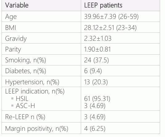
The average age of 64 patients who underwent LEEP was 39.96±7.39 years. The mean BMI of patients was 28.12±2.51. The mean gravidy and parity of those patients were 2.32±1.03 and 1.90±0.81, respectively. In 64 patients, 37.5% have smoking, 9.4% have diabetes mellitus and 20.3% have hypertension history. Indications for LEEP were HSIL in 61 patients and ASC-H in 3 patients.
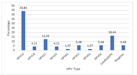
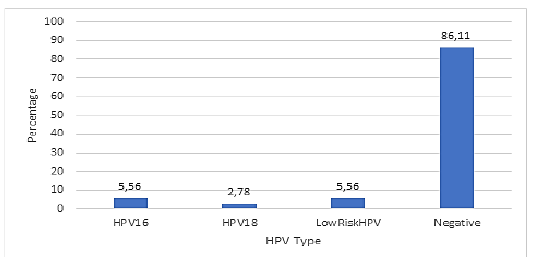
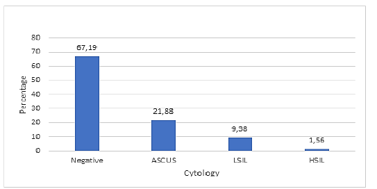
Distribution of HPV types of patients before LEEP are shown in Figure 1. The most common detected HPV type was HPV 16 (43.84%) before LEEP. HPV 31 was the second common high risk HPV type seen before LEEP. Of the 4 patients, there was no HPV type detected before LEEP. Distribution of HPV types of patients after two years follow-up for LEEP are shown in Figure 2. Of the 35 patients, 86.11 % of patients was negative for HPV after two years follow-up. The most common detected HPV types were HPV 16, low risk HPV types and HPV 18, respectively. The cytological outcomes outcomes of the study group after two years follow-up were documented in Figure 3. 67.19% of patients who underwent LEEP have negative cytology after two years follow-up. 21.88% of patients have ASCUS, 9.38% of the patients have LSIL and 1.56 % of patients have HSIL cytology after two years follow-up. Four of 64 LEEP specimen (6.25%), surgical margin status were positive. There were 3 patients who underwent re-LEEP in two years period for HSIL. All of the patients who underwent re-LEEP were positive in terms of surgical margin status. One patient of the studied group underwent bilateral salpingo opherectomy laparoscopic hysterectomy and for abnormal uterine bleeding in the sixth month of follow-up period.
Discussion
In this research, we evaluated the two years follow-up outcomes of patients who underwent LEEP procedure for HSIL/ASC-H proven histo pathologically. According to this report, it was revealed that LEEP procedure was an effective surgical option in the management of high grade cervical preinvasive diseases. Indeed, there were detected satisfactory outcomes and regression in cytopathological and HPV status of patients after two years follow-up period of LEEP.
In the present study, 1.56% of patients with pre-operative CIN 2/3 in colposcopic biopsy showed persistance of the CIN 2/3 lesion. We observed resolution of the disease in 67.19% of patients with preoperative CIN 2/3. However, nearly 98% of the patients showed regression after LEEP. There was a dramatic regression after LEEP in two years follow-up period. Our study demonstrates a very high efficacy of LEEP for the treatment of CIN 2/3 with persistent disease in only 1.56% of patients and no recurrence in two year follow-up period. Previously published recurrence rates after LEEP procedure were 1.5, 2.2, 9.1 and 11.9% for CIN 2/3 (6–9). According to the results of these studies, there were much more margin positive cases than those in our study. We think that low margin positive cases may have a major role in lower recurrence rate of our study.
Papalia and his colleagues used laser ablation in combination with LEEP and reported lower recurrence rates as 3.2% (10). In our hospital, we did not have laser ablation technology. Although we did not used laser in this study, recurrence was not seen in any patient.
After LEEP operations, follow-up of HPV status of patients is very important. Kim et al reported 85% of patients after LEEP showed HPV clearance in the six month follow-up (11). Jing et al. reported HPV positivity of the patients as 95.6% before LEEP and 29.8% after LEEP in two years follow-up period (12). Bae et al. reported HPV status as 93% before and as 35.1% after LEEP and they concluded that detection of HPV status after conizaiton may help to identify treatment failure or recurrence (13). In our study, we observed that the ratios of HPV negative patients increased from 5% to 86% after LEEP. Although the follow-up time was longer as two years, HPV clearance rate was similar to Kim et al.’s study. In this context, our study conforms that HPV clearance rate increases gradually after LEEP. But the strict follow-up of HPV status of patients is necessary after operation for HPV persistence, especially for high risk genotypes. Some authors had reported that high-risk HPV status after LEEP is a better predictor of CIN recurrence than colposcopy or cytology results (11, 14). However, some authors stated that high-risk HPV load prior to conization serves as a causative agent for recurrence(9).
In the study of Jing et al., 71% of patients who underwent LEEP were surgical margin positive (12). In our study, only 4 (6.25%) patients were positive in regard of surgical margin. Surgical margin status of LEEP patients is a major risk factor for recurrence or persistence of cervical intraepithelial neoplasia (15). Patients with positive surgical margin after LEEP have more likely experience recurrent CIN (16). In the literature, there are recurrence rates as high as 47% for patients with positive margin (17). In our study population, three of 4 patients with positive margin underwent LEEP again. Of these 3 patients, all of them were positive for HPV 16 subtype. Spontaneous regression was seen in one patient and HPV clearance was detected in her follow-up period. These findings of our study emphasizes the critical importance of persistence in high-risk HPV subtypes especially for HPV 16 at the follow up period of margin positive cases. Moreover, Bruno et al. reported that 40% of the relapsing cases in their study were seen in margin negative cases with persistent HPV 16 infection (18). We strongly agree with this opinion. If HPV 16 persistency is seen at the follow-up, a crucial attention must be given even the pathology report is margin negative.
The limitations of our study were having the retrospective design and small sampling size with lack of a comparative group. The strength of our study was the completeness of cytological data and the low lost-to-follow-up rate. In future, researches may be planned as in prospective nature designed to evaluate adverse events, oncological findings and the quality of life of patients.
CIN recurrency may be encountered mostly within the subsequent two years follow-up period of LEEP. The rate of recurrence after LEEP is higher in HPV positive. Therefore, these patients necessitates careful monitorization. In identification of residual lesions or recurrencies, Pap-smear and HPV testing have a central role at the follow-up strategies. However, recommendation favoring the use of more effective follow-up strategies for the margin positive patients should be supported with researches involving longer follow-up periods and larger patient series.
Declarations
- Author contributions (CRediT)
- All authors contributed equally to all 14 CRediT roles (Conceptualization, Data curation, Formal analysis, Funding acquisition, Investigation, Methodology, Project administration, Resources, Software, Supervision, Validation, Visualization, Writing – original draft, Writing – review & editing).
- Funding
-
Funding information was not reported in the published version of this article.
- Conflicts of interest
- The author declared no conflicts of interests.
- Ethics approval
- This retrospective study was approved by the local ethics committee for clinical research of Mugla Sitki Kocman University, Faculty of Medicine, Mugla, Turkey.
- Data availability
- A data availability statement was not reported in the published version of this article.
References (18)
14 of 18 references are linked to their DOI.
- Rositch AF, Soeters HM, Offutt-Powell TN, Wheeler BS, Taylor SM, Smith JS. The incidence of human papillomavirus infection following treatment for cervical neoplasia: a systematic review. Gynecol Oncol. 2014;132:767-779 DOI Crossref PubMed Google Scholar
- Hoffman SR, Le T, Lockhart A, Sanusi A, Dal Santo L, Davis M, et al. Patterns of persistent HPV infection after treatment for cervical intraepithelial neoplasia (CIN): A systematic review. Int J Cancer. 2017;141:8-23 DOI Crossref PubMed Google Scholar
- Chelmow D. Committee on Practice Bulletins-Gynecology. Practice Bulletin No. 168: Cervical Cancer Screening and Prevention. Obstet Gynecol. 2016;128:e111-e130 PubMed Google Scholar
- Santesso N, Mustafa RA, Wiercioch W, Kehar R, Gandhi S, Chen Y, et al. Systematic reviews and meta-analyses of benefits and harms of cryotherapy, LEEP, and cold knife Loop Electrosurgical Excision Procedure Burak Sezgin conization to treat cervical intraepithelial neoplasia. Int J Gynecol Obstet. 2016;132:266 –71 DOI Crossref PubMed Google Scholar
- Chen LM, Liu L, Tao X, He Y, Guo LP, Zhang HW, et al. Analysis of recurrence and its influencing factors in patients with cervical HSIL within 24 months after LEEP. Zhonghua Fu Chan Ke Za Zhi. 2019;54:534–40 PubMed Google Scholar
- Duesing N, Schwarz J, Choschzick M, Jaenicke F, Gieseking F, Issa R, et al. Assessment of cervical intraepithelial neoplasia (CIN) with colposcopic biopsy and efficacy of loop electrosurgical excision procedure (LEEP). Arch Gynecol Obstet. 2012;286:1549–54 DOI Crossref PubMed Google Scholar
- Cecchini S, Visioli CB, Zappa M, Ciatto S. Recurrence after treatment by loop electrosurgical excision procedure (LEEP) of high-grade cervical intraepithelial neoplasia. Tumori. 2002;88:478-480 DOI Crossref PubMed Google Scholar
- Fan Q, Tay SK, Shen K. Loop electrosurgical excision procedure: a valuable method for the treatment of cervical intraepithelial neoplasia. Zhonghua Fu Chan Ke Za Zhi. 2001;36:271-4 PubMed Google Scholar
- Alonso I, Torné A, Puig-Tintoré LM, Esteve R, Quinto L, Campo E, et al. Pre- and post-conization high-risk HPV testing predicts residual/recurrent disease in patients treated for CIN 2-3. Gynecol Oncol. 2006;103:631–6 DOI Crossref PubMed Google Scholar
- Papalia N, Rohla A, Tang S, Nation J, Nelson G. Defining the short-term disease recurrence after loop electrosurgical excision procedure (LEEP). BMC Womens Health. 2020; 20(1): DOI Crossref PubMed Google Scholar
- Young TK, Lee JM, Hur SY, Cho CH, Kim YT, Seung CK, et al. Clearance of human papillomavirus infection after successful conization in patients with cervical intraepithelial neoplasia. Int J Cancer. 2010;126:1903– 9 DOI Crossref PubMed Google Scholar
- Jing L, Dan W, Zhunan L, Ying X, Yi C. Residual lesions in uterine specimens after loop electrosurgical excision procedure in patients with CIN. Arch Gynecol Obstet 2018; 298:805– 12. DOI Crossref PubMed Google Scholar
- Bae JH, Kim CJ, Park TC, Namkoong SE, Park JS. Persistence of human papillomavirus as a predictor for treatment failure after loop electrosurgical excision procedure. Int J Gynecol Cancer 2007;17:1271–7 DOI Crossref PubMed Google Scholar
- Gallwas J, Ditsch N, Hillemanns P, Friese K, Thaler C, Dannecker C. The significance of HPV in the follow-up period after treatment for CIN. Eur J Gynaecol Oncol. 2010;31:27-30 PubMed Google Scholar
- Bornstein J, Schwartz J, Perri A, Harroch J, Zarfati D. Tools for post LEEP surveillance. Obstet Gynecol Surv. 2004;59:663-8 DOI Crossref PubMed Google Scholar
- Shafi MI, Dunn JA, Buxton EJ, Finn CB, Jordan JA, Luesley DM. Abnormal cervical cytology following large loop excision of the transformation zone: a case controlled study. Br J Obstet Gynaecol. 1993;100:145-148 DOI Crossref PubMed Google Scholar
- Bornstein J, Yaakov Z, Pascal B, Faktor J, Baram A, Zarfati D, et al. Decision-making in the colposcopy clinic - A critical analysis. Eur J Obstet Gynecol Reprod Biol. 1999;85:219–24 DOI Crossref PubMed Google Scholar
- Bruno MT, Cassaro N, Garofalo S, Boemi S. HPV16 persistent infection and recurrent disease after LEEP. Virol J. 2019;16:148. DOI Crossref PubMed Google Scholar
Respond to this article
Comments on a published article are welcome as a Letter to the Editor.
Get an e-mail when new articles in your fields are published
Questions to the authors
Questions are moderated: an editor approves each question, the authors answer, and an editor approves the answer before both appear here.
No answered questions yet.
Ask the authors a question
Discussion
Comments are moderated: they appear after the writer confirms the e-mail address and an editor approves them. Each comment carries a conflict-of-interest statement.
No comments yet.
Write a comment
Read the full text
Open the PDF here
Loading the PDF…
If the PDF does not show here, use the download button above.
How to cite this article
Sezgin B. Outcomes of Two Years Follow-Up after Loop Electrosurgical Excision Procedure in a University Hospital. Ulutas Med J. 2020;6(3):156-161. doi:10.5455/umj.20200624063656
Sezgin, B. (2020). Outcomes of Two Years Follow-Up after Loop Electrosurgical Excision Procedure in a University Hospital. The Ulutas Medical Journal, 6(3), 156–161. https://doi.org/10.5455/umj.20200624063656
Sezgin, B. (2020) 'Outcomes of Two Years Follow-Up after Loop Electrosurgical Excision Procedure in a University Hospital', The Ulutas Medical Journal, 6(3), pp. 156–161. doi: 10.5455/umj.20200624063656.
@article{umj118487,
  title = {Outcomes of Two Years Follow-Up after Loop Electrosurgical Excision Procedure in a University Hospital},
  author = {Sezgin, Burak},
  journal = {The Ulutas Medical Journal},
  year = {2020},
  volume = {6},
  number = {3},
  pages = {156--161},
  doi = {10.5455/umj.20200624063656},
  url = {https://www.ulutasmedicaljournal.com/?mno=118487},
  issn = {2149-388X}
}