The Ulutas Medical Journal · ISSN 2149-0430 (print), 2149-388X (online) · https://www.ulutasmedicaljournal.com/?mno=141168
Original Article · Open access
The Role of Hysterectomy in the Occurrence of Postmenopausal Osteoarthritis in Rats Undergoing Hysterectomy, Oophorectomy and Oophorectomy
Also available in: العربية · Türkçe · 中文
Abstract
- Introduction
- Hysterectomy is the most common gynecological operation performed after cesarean operation. This animal study investigates the role of hysterectomy on the development of postmenopausal osteoarthritis through surgical approaches.
- Materials and Methods
- The study sample comprised 40 mature female Albino rats that were randomly separated into 4 surgical groups of 10 as control, oophorectomy, hysterectomy, and ovariohysterectomy. Serial sections were taken for histological examination. The average scores of each observer were calculated and used in analyses.
- Results
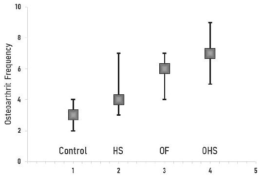
- When the osteoarthritis scores were compared according to the groups, the highest score was found in the ovariohysterectomy group, followed by oophorectomy, hysterectomy, and control groups, respectively. The difference between the groups in respect of osteoarthritis was found to be significant (p<0.0001). When the osteoarthritis scores of the groups were compared in pairs, significant differences were determined between the hysterectomy and the oophorectomy (p<0.05), between the hysterectomy and the ovariohysterectomy (p<0.01), and between the hysterectomy, the oophorectomy, and the ovariohysterectomy and the control groups (p<0.05, p<0.05, p<0.001, respectively).
- Conclusion
- A higher osteoarthritis score in the ovariohysterectomy compared to the oophorectomy concluded that hysterectomy caused the development of osteoarthritis in both the groups in which the ovaries were protected and not protected.
Keywords
Full text
Introduction
Hysterectomy is very common gynecological operation performed after cesarean and is applied to women aged 30-54 years (1). From an examination of indications, the rate of cancer-related operations has been reported to be approximately 11.2% (2). The most common operation accompanying a hysterectomy is oophorectomy, that most of the hysterectomies are applied to patients between the ages of 30 and 54 years and approximately 10% of the operations are due to cancer, it is important to decide whether the ovaries can be preserved or not, to protect the patient from the negative effects of surgical menopause (3, 4).
There is a general belief among gynecologists that patients with ovarian protection are spared the negative effects of surgical menopause (5). In contrary, in various epidemiological and experimental studies, although the ovaries are preserved (6), it was reported that hysterectomy alone causes a decrease in ovarian reserve, osteoarthritis due to cardiovascular thrombo embolism, menopause, and osteoporosis (3). Menopause, which is another name for the natural decrease in the functions of the ovaries, is the permanent loss of menstruation as a result of the loss of ovarian follicular activity (7). With the increase in overall life expectancy over the past century, more than one-third of women's lives constitute postmenopausal years (8). Joint complaints, one of the most important menopausal symptoms, are seen in more than 50% of postmenopausal women (9).
Osteoarthritis is a disorder characterized by progressive degeneration of the articular cartilage, remodelling in the subchondral bone, marginal osteophyte formation, and loss of mechanical function (10). Osteoarthritis is seen in the knee joint in 6% of the population over 30 years in the United States, and in the hip joint in 3% (11). While osteoarthritis is more common in men aged <50 than women, the incidence of osteoarthritis increases in menopausal women (4). The prevalence of osteoarthritis decreases significantly in women who receive hormone replacement (6). In a study by Andersen et al. a postmenopausal osteoarthritis was created in rats with oophorectomy and it was reported that surgical menopause is a process that significantly accelerates the development of osteoarthritis (12).
The aim of this study was to investigate the role of hysterectomy on the development of osteoarthritis, which is an important morbidity factor for postmenopausal women, through the evaluation of a rat model of hysterectomy, ovariohystectomy and oophorectomy groups.
Materials and Methods
Ethical Statement
This animal study was conducted in Cumhuriyet University, Faculty of Medicine, Animal Trial Laboratory in collaboration with the Pathology. The study was approved by Experimental Animals Ethics Committee of Cumhuriyet University (decision number 0001042004-22).
Study Design
The study sample comprised 40 mature female Wistar Albino rats, aged 6 months, and each weighing 200-250 g. Before the study, the rats were brought to the specified weight by keeping them under standard laboratory conditions. Feed and water were maintained twice a day, without any quantity limitations. The 40 rats were randomly separated into 4 groups of 10 as control, oophorectomy, hysterectomy and ovariohystectomy groups.
Surgical Application
All the rats were administered 3mg/kg xylazine HCl (Rompun, Bayer, Istanbul) and 3mg/kg ketamine HCI (Pfizer, Istanbul) intramuscularly. The rats were laparotomized with a midline incision of 3 cm, starting just above the external urethral orifice. Only laparotomy was performed on rats in the control group to exclude the effects of anesthetics and surgical procedure. Following the laparotomy; i) In the control, no additional procedure was made and the peritoneum and muscle layers were closed continuously using 3/0 polyglactin silt (Vicryl, Ethicon, Jonson-Jonson Intl. Stevens- Woluwe, BELGIUM) and for the skin, 4/0.
ii) In the hysterectomy group, as an initial hysterectomy procedure, the utero-ovarian artery and vein were sutured with 4/0 absorbable polyglalctin to protect ovarian blood flow. Both uterine tubes were removed from the junction with the uterina to the level of the cervix. The peritoneum and muscle were closed by 3/0, skin with 4/0 polyglactin, and continuous silts.
iii) In the oophorectomy, the infidubulopelvic ligament connecting the ovary to the posterior abdominal wall, the suspensor ligament connecting the uterine horns, the ovarian artery and the vein were held with a clamp, tied with a single suture and then cut and oophorectomy was performed. Following the oophorectomy, the peritoneum and muscle layers were closed with 3/0 and the skin with 4/0 polyglactin using continuous sutures.
iv) In the ovariohystectomy group, suspensor ligaments connecting the ovary to the posterior abdominal wall to the infindubulopelvic and uterine organs were tied to the ovarian artery and vein. Then, the cardinal ligaments, uterine artery and vein fixing the uterine horn, were cut and tied. Uterine booms were removed by cutting at the level of cervix. The peritoneum and muscle layers were closed using 3/0 and the skin 4/0 polyglactin with continuous sutures. While no complications related to the operation were observed in the early period, the rats were kept in the standard laboratory environment for 6 months. At the end of six months, all rats were euthanised with the cervical dislocation method under anesthesia. The knee joint was removed, including the distal 1 cm of the femur and the proximal 1 cm of the tibia. Surrounding muscle tissues were carefully excised without damaging the joint integrity. The joints were fixed in 10% formaldehyde (40% Formaldehyde, Carlo Erba Reagenti SpA, Milano, ITALY) solution, 10% HCl (Hydrochloric Acid, Kimetsan, Ankara) and 90% formic acid (HCOOH, Kimetsan, Ankara) for 3 weeks. After confirmation that decalcification process was completed, the routine follow-up procedure was applied to the samples taken from the tissues to show the bone ends and other structures constituting the knee joint, and they were embedded in paraffin blocks.
Histopathological Assessment
Serial sections were taken for a histological examination. All sections were stained with hematoxylin-eosin for examination of cellularity and toluidine blue for investigation of proteo glycan content. These sections were examined under a light microscope (Nilcon Eclipse E600, Nikon Instech Co., JAPAN). In the evaluation of histopathological changes of osteoarthritis, the modified Galois et al. A scoring system was used(16). Accordingly, joint structure, cellularity, staining with toluidine blue, thickness of the hypertrophic chondrocyte layer, osteolysis and remodelling in the bone were taken into consideration. Histological examination was performed separately by two pathologists blinded to the groups to which the sections belonged. The average scores of each observer were calculated and used in analyses.
Statistical Evaluation
Bioistatistical analysis of the patients’ data were analyzed by SPSS software (version 23- Windows). The Kruskal-Wallis ANOVA test was applied to evaluate the significance of the data obtained from the groups. The Student- Newman-Keuls test was applied as a post hoc test to show whether there was a significant difference between the groups. The data are presented as median (min-max) values. A value of p<0.05 was considered significant.
Results
The present study was completed without postoperative loss in all the groups. When the osteoarthritis scores were compared according to the groups, the highest score was found in the ovariohysterectomy group, followed by oophorectomy, hysterectomy and control groups, respectively. The median values of the osteoarthritis scores of the groups were at most 7 (range: 5-9) in the ovariohysterectomy. In terms of osteoarthritis score, the highest scores of 6 (range: 4-7) were in the rats that underwent oophorectomy after the ovariohysterectomy. Scores of 4 (range: 3-7) and 3 (range: 2-4) were found in the hysterectomy and control group. The difference between the groups in respect of osteoarthritis was significant (p<0.0001). When the osteoarthritis scores were compared in pairs, significant difference was determined between the hysterectomy and oophorectomy groups(p<0.05). There was a strong significance between hysterectomy and ovariohysterectomy group (p<0.01). Also we found relationship between the hysterectomy, the oophorectomy, the ovariohysterectomy and control groups (p<0.05, p<0.05,p<0.001,respectively)(Figure-1).
Discussion
Although many factors are held responsible for the development of osteoarthritis, the ways in which these factors play a role in the pathogenesis have not been clarified yet. In this rat model study, as a suitable species on which to create an osteoarthritis model, the results have proven that hysterectomy is significantly associated with osteoarthritis. This supports the theory that estrogen levels are of great importance in the development of osteoarthritis in females. In this animal experiment, the surgical removal of the female reproductive organs was seen to have a significant impact on triggering the development of osteoarthritis through the estrogen level affected by the aggressive surgical approach.
Although there has been much discussion about osteoarthritis-related etiological factors, it is accepted that there is a multifactorial pathology in which biomechanical, genetic and metabolic factors play a role (11). The increasing prevalence of osteoarthritis among women from around the age of 50 years suggests that the hormonal changes observed in menopause have an effect on osteoarthritis (6). As a result of studies showing that the presence of estrogen receptors in cartilage tissue and hormone replacement therapy reduce the prevalence of osteoarthritis, it has been proven that estrogen plays a role in the development of osteoarthritis. Many scoring systems have been developed by different researchers for grading histopathological changes due to osteoarthritis. In the current study, the Mankin scoring system modified by Galois et al was used (13). Histopathological examination was performed by two different pathologists blinded to the study. In a study by Ostergaard et al. histopathological changes in normal and osteoarthritic knee joints were scored twice at a 1-week interval by three different pathologists blinded to the groups. The intra-observer agreement was reported as 22% - 33%, and the inter-observer agreement as 13% - 25% (14). In the current study, the arithmetic mean of the scores obtained from both observers was used to minimise the effect of inter-observer differences on the results.
The correlation between hysterectomy and osteoarthritis is a controversial issue. In the current study, the osteoarthritis score in the hysterectomy group was significantly higher than that of the control group (p <0.05). Contrary to these findings, there are studies in literature that have reported that hysterectomy is not associated with the development of osteoarthritis or is protective against osteo arthritis. Stöve et al. evaluated 504 women scheduled for arthroplasty, and determined no significant relationship between osteoarthritis and hysterectomy in patient who emerged hysterectomy, while hysterectomy was found to have a significant protective effect for the generalized form of the disease (15). However, in that study, the rate of use of hormone replacement therapy in hysterectomy history was 16%, and 10% in patients without hysterectomy. In their Osteo porotic Fracture Study, Hochberg et al. Found that surgical menopause was protective against osteoarthritis, which was attributed to the higher rate of hormone replacement therapy use in those who had hysterectomy (16). Cooley et al. reported no connection between hand osteoarthritis and hysterectomy and Erb et al. found no relationship between hysterectomy and osteoarthritis (17). However, there are also articles in the literature that support the current study findings. In another study of 97 severe osteoarthritis cases, a statistically significant relationship was found between hysterectomy and osteoarthritis, similar to our findings (4). Of the additional operations performed with hysterectomy, oophorectomy is the most common (18). Despite the general belief among gynecologists that the protection of the ovaries protects the patient from the pathologies associated with menopause and menopause (5), it has been proven that hysterectomy affects ovarian functions and increases the risk of early menopause (19). It has been claimed in various studies that there is a decrease in ovarian functions and early ovarian insufficiency after hysterectomy (20). The reason is that hysterectomy reduces ovarian blood flow, the paracrine effect of the uterus on the ovary, or the endocrine effect on other tissues (21). In the current study, the osteoarthritis score of the ovariohysterectomy group was significantly higher than that of the oophorectomy group. In the ovariohysterectomy group, it was noted that the hysterectomy performed in addition to the oophorectomy made a significant difference in respect of the paracrine effect of the uterus on the ovarian tissue and the effects of hysterectomy on ovarian blood flow, in addition to the endocrine functions of the uterus. Consistent with this view, the uterus produces various cytokines and growth factors, independent of estrogen level changes, and endocrine effects that stimulate osteoblasts and bone cells have been found (22–24).
In the current study, the scores obtained from the oophorectomy group were higher than those of the control group, demonstrating that the continuation of ovarian functions is protective against osteoarthritis, consistent with many studies in the literature. In this context, osteoarthritis scores are expected to be lower in cases where the ovaries are preserved during hysterectomy compared to cases without preservation. That higher scores were obtained in the ovariohysterectomy group compared to the hysterectomy group was found to be consistent with the aforementioned view. In contrast, Spector et al. reported that the risk of osteoarthritis was increased in women who had undergone hysterectomy, but oophorectomy did not create any additional risk (25). The average osteoarthritis score obtained in the current study oophorectomy group was significantly higher than that of the control group. Hoegh-Andersen et al conducted a study on female rats that examined the effects of oophorectomy on the development of osteoarthritis, similar to the current study model, and the osteoarthritis scores were significantly higher in the oophorectomy group, consistent with the current study results(12). In this study, an answer was sought to the question of what age rats should be used to create an osteoarthritis model. As a result of the measurement of collagen type-II degradation products in female and male rats, which is a metabolite originating from cartilage tissue during normal development of the skeletal system, it has been reported that the level of this metabolite decreases with age and this decrease is observed in the third month at most. In the light of this information, it has been suggested to use rats older than 3 months in models to be created for osteoarthritis. Since the 6-month old rats used in the current study had completed normal development and reached skeletal system maturity, there was considered to be no interference from joint changes due to growth and development. There are human and animal studies in the literature examining the factors that contribute to the development of osteoarthritis (10, 25–27). One of the advantages that animal models have over human studies is that they allow for detailed investigation in the diagnosis of the disease. While histopathological examination can be made in animals by direct dissection of the joints, this is only possible in humans when applying prosthesis, in postmortem biopsies, or in cases with severe joint damage (11, 28). Rupture of the meniscus and cutting the ligaments have been used for this purpose in various studies to create an osteoarthritis model However, osteoarthritis caused by these methods does not allow early stage changes, but only the advanced stage changes in which the joint structure shows severe functional and structural impairment. The advantage of osteoarthrit model obtained by oophorectomy is that the more common mild and moderate changes that occur in humans can be observed. Rats were selected for this study because of their genetic homogeneity, low cost and easy availability, short cycle and similarity to the human skeletal system. There are studies in the literature with rats and other members of the rodents, such as hamsters and mice (12, 13). There were limitations to this experimental rat model study, primarily that there was no opportunity to make a mechanical evaluation of osteoarthritis as in human subjects, and that only the short-term effects could be evaluated.
Conclusion
Using rats to create an osteoarthritis model, the effect of hysterectomy on the development of osteoarthritis was investigated. Of the species that can be used to examine the histopathological changes due to osteoarthritis, the use of rats is a suitable choice because of the low cost and the skeletal system similarity to humans. The osteoarthritis score in the hysterectomy group rats was found to be significantly higher than that of the control group. A significantly higher osteoarthritis score was found in the ovariohysterectomy group compared to the oophorectomy group. Thus it can be concluded that hysterectomy caused the development of osteoarthritis in both the groups in which the ovaries were protected and the ovaries were not protected.
Declarations
- Author contributions (CRediT)
- All authors contributed equally to all 14 CRediT roles (Conceptualization, Data curation, Formal analysis, Funding acquisition, Investigation, Methodology, Project administration, Resources, Software, Supervision, Validation, Visualization, Writing – original draft, Writing – review & editing).
- Funding
-
Conflict of Interest The authors declare that there is no conflict of interest and no financial support for the study.
- Conflicts of interest
- The authors declare that there is no conflict of interest and no financial support for the study.
- Ethics approval
- The study was approved by Experimental Animals Ethics Committee of Cumhuriyet University (decision number 0001042004-22).
- Data availability
- A data availability statement was not reported in the published version of this article.
References (28)
25 of 28 references are linked to their DOI.
- Atay V, Ceyhan T, Baser I, Gungor S, Goktolga U, Muhcu M. Hysterectomy with preservation of both ovaries does not result in premature ovarian failure. The Journal of international medical research. 2007;35(3):416-21 DOI Crossref PubMed Google Scholar
- Kuppermann M, Varner RE, Summitt RL, Jr., Learman LA, Ireland C, Vittinghoff E, et al. Effect of hysterectomy vs medical treatment on health-related quality of life and sexual functioning: the medicine or surgery (Ms) randomized trial. Jama. 2004;291(12):1447-55 DOI Crossref PubMed Google Scholar
- Uccella S, Capozzi VA, Ricco M, Perrone E, Zanello M, Ferrari S, et al. Sexual Function following Laparoscopic versus Trans vaginal Closure of the Vaginal Vault after Laparoscopic Hysterectomy: Secondary Analysis of a Randomized Trial by the Italian Society of Gynecological Endoscopy Using a Validated Questionnaire. Journal of minimally invasive gynecology. 2020;27(1):186-94 DOI Crossref PubMed Google Scholar
- de Klerk BM, Schiphof D, Groeneveld FP, Koes BW, van Osch GJ, van Meurs JB, et al. Limited evidence for a protective effect of unopposed oestrogen therapy for osteoarthritis of the hip: systematic review. Rheumatology. 2009;48(2):104-12 DOI Crossref PubMed Google Scholar
- Gokmen Karasu AF, Ates S, Gurbuz T, Sahin N, Takmaz T, Aydin S. A Clinico-pathological Study of Transvaginal Endometrial Thickness Measurement in Asymptomatic Post menopausal Patients and Patients with Postmenopausal Bleeding. Gynecology Obstetrics; Reproductive Medicine. 2019;25(2):85-8 DOI Crossref PubMed Google Scholar
- Kermode-Scott B. Ineffective surgical procedures are used to treat Canadians with osteoarthritis. BMJ (Clinical research ed). 2010;341:c7436 DOI Crossref PubMed Google Scholar
- Lowery AJ, Sweeney KJ, Molloy AP, Hennessy E, Curran C, Kerin MJ. The effect of menopause and hysterectomy on systemic vascular endothelial growth factor in women under going surgery for breast cancer. BMC cancer. 2008;8:279 DOI Crossref PubMed Google Scholar
- Van Lieshout LAM, Pijlman B, Vos MC, de Groot MJM, Houterman S, Coppus S, et al. Opportunistic salpingectomy in women undergoing hysterectomy: Results from the HYSTUB randomised controlled trial. Maturitas. 2018;107:1-6 DOI Crossref PubMed Google Scholar
- Findley AD, Siedhoff MT, Hobbs KA, Steege JF, Carey ET, McCall CA, et al. Short-term effects of salpingectomy during laparoscopic hysterectomy on ovarian reserve: a pilot randomized controlled trial. Fertility and sterility. 2013;100 (6):1704-8 36 77 Hysterectomy in Postmenopausal Osteoarthritis Orhan Yanar DOI Crossref PubMed Google Scholar
- Wang A, Zawadzki N, Hedlin H, LeBlanc E, Budrys N, Van Horn L, et al. Reproductive history and osteoarthritis in the Women's Health Initiative. Scandinavian journal of rheumatology. 2021;50(1):58-67 DOI Crossref PubMed Google Scholar
- Fontana L, Neel S, Claise JM, Ughetto S, Catilina P. Osteoarthritis of the thumb carpometacarpal joint in women and occupational risk factors: a case-control study. The Journal of hand surgery. 2007;32(4):459-65 DOI Crossref PubMed Google Scholar
- Høegh-Andersen P, Tankó LB, Andersen TL, Lundberg CV, Mo JA, Heegaard AM, et al. Ovariectomized rats as a model of postmenopausal osteoarthritis: validation and application. Arthritis research & therapy. 2004;6(2):R169-80 DOI Crossref PubMed Google Scholar
- Galois L, Etienne S, Grossin L, Watrin-Pinzano A, Cournil-Henrionnet C, Loeuille D, et al. Dose-response relationship for exercise on severity of experimental osteoarthritis in rats: a pilot study. Osteoarthritis and cartilage. 2004;12(10):779-86. DOI Crossref PubMed Google Scholar
- Ostergaard M, Halberg P. [Intra-articular glucocorticoid injections in joint diseases]. Ugeskrift for laeger. 1999;161(5): 582-6 PubMed Google Scholar
- Stöve J, Stürmer T, Kessler S, Brenner H, Puhl W, Günther KP. Hysterectomy and patterns of osteoarthritis. The Ulm Osteoarthritis Study. Scandinavian journal of rheumatology. 2001; 30(6):340-5 PubMed Google Scholar
- Hochberg MC, Altman RD, April KT, Benkhalti M, Guyatt G, McGowan J, et al. American College of Rheumatology 2012 recommendations for the use of nonpharmacologic and pharmacologic therapies in osteoarthritis of the hand, hip, and knee. Arthritis care & research. 2012;64(4):465-74 DOI Crossref PubMed Google Scholar
- Cooley HM, Stankovich J, Jones G. The association between hormonal and reproductive factors and hand osteoarthritis. Maturitas. 2003;45(4):257-65 DOI Crossref PubMed Google Scholar
- Manson JE, Aragaki AK, Bassuk SS, Chlebowski RT, Anderson GL, Rossouw JE, et al. Menopausal Estrogen-Alone Therapy and Health Outcomes in Women With and Without Bilateral Oophorectomy: A Randomized Trial. Annals of internal medicine. 2019;171(6):406-14 DOI Crossref PubMed Google Scholar
- Siyam T, Ross S, Shandro T, Hagen S, Battochio L, Yuksel N. Perspectives and decision-making about menopausal therapies in women who had bilateral oophorectomy. Menopause (New York, NY). 2018;25(7):795-802 DOI Crossref PubMed Google Scholar
- Luo J, Manson JE, Urrutia RP, Hendryx M, LeBlanc ES, Margolis KL. Risk of Diabetes After Hysterectomy With or Without Oophorectomy in Postmenopausal Women. American journal of epidemiology. 2017;185(9):777-85 DOI Crossref PubMed Google Scholar
- Chen L, Mishra GD, Dobson AJ, Wilson LF, Jones MA. Protective effect of hormone therapy among women with hysterectomy/oophorectomy. Human reproduction (Oxford, England). 2017;32(4):885-92 DOI Crossref PubMed Google Scholar
- Stanczyk FZ, Chaikittisilpa S, Sriprasert I, Rafatnia A, Nadadur M, Mishell DR, Jr. Circulating androgen levels before and after oophorectomy in premenopausal and postmenopausal women. Climacteric: the journal of the International Menopause Society. 2019;22(2):169-74 DOI Crossref PubMed Google Scholar
- Harvey LFB, Abramson VG, Alvarez J, DeStephano C, Hur HC, Lee K, et al. Surgical Findings and Outcomes in Premenopausal Breast Cancer Patients Undergoing Oophorectomy: A Multicenter Review From the Society of Gynecologic Surgeons Fellows Pelvic Research Network. Journal of minimally invasive gynecology. 2018;25(1):111-5 DOI Crossref PubMed Google Scholar
- Wong J, Murji A, Sunderji Z, Chow O, Shapiro J, Wolfman W, et al. Unnecessary bilateral salpingo-oophorectomy at the time of hysterectomy and potential for ovarian preservation. Menopause (New York, NY). 2020;28(1):8-11 DOI Crossref PubMed Google Scholar
- Spector TD, Hart DJ, Brown P, Almeyda J, Dacre JE, Doyle DV, et al. Frequency of osteoarthritis in hysterectomized women. The Journal of rheumatology. 1991;18(12):1877-83 PubMed Google Scholar
- Kroon FPB, Kortekaas MC, Boonen A, Böhringer S, Reijnierse M, Rosendaal FR, et al. Results of a 6-week treatment with 10 mg prednisolone in patients with hand osteoarthritis (HOPE): a double-blind, randomised, placebo-controlled trial. Lancet. 2019;394(10213):1993-2001 DOI Crossref PubMed Google Scholar
- Spector TD, Nandra D, Hart DJ, Doyle DV. Is hormone replacement therapy protective for hand and knee osteoa rthritis in women?: The Chingford Study. Annals of the rheumatic diseases. 1997;56(7):432-4 DOI Crossref PubMed Google Scholar
- Chlebowski RT, Cirillo DJ, Eaton CB, Stefanick ML, Pettinger M, Carbone LD, et al. Estrogen alone and joint symptoms in the Women's Health Initiative randomized trial. Menopause. 2018;25(11):1313-20 DOI Crossref PubMed Google Scholar
Respond to this article
Comments on a published article are welcome as a Letter to the Editor.
Get an e-mail when new articles in your fields are published
Questions to the authors
Questions are moderated: an editor approves each question, the authors answer, and an editor approves the answer before both appear here.
No answered questions yet.
Ask the authors a question
Discussion
Comments are moderated: they appear after the writer confirms the e-mail address and an editor approves them. Each comment carries a conflict-of-interest statement.
No comments yet.
Write a comment
Tell me when this article is cited
Leave your e-mail address and confirm it. These notices are not being sent yet; your confirmed address is kept for them and used for nothing else.
Read the full text
Open the PDF here
Loading the PDF…
If the PDF does not show here, use the download button above.
How to cite this article
Yanar O. The Role of Hysterectomy in the Occurrence of Postmenopausal Osteoarthritis in Rats Undergoing Hysterectomy, Oophorectomy and Oophorectomy. Ulutas Med J. 2022;8(1):30-36. doi:10.5455/umj.20220110060638
Yanar, O. (2022). The Role of Hysterectomy in the Occurrence of Postmenopausal Osteoarthritis in Rats Undergoing Hysterectomy, Oophorectomy and Oophorectomy. The Ulutas Medical Journal, 8(1), 30–36. https://doi.org/10.5455/umj.20220110060638
Yanar, O. (2022) 'The Role of Hysterectomy in the Occurrence of Postmenopausal Osteoarthritis in Rats Undergoing Hysterectomy, Oophorectomy and Oophorectomy', The Ulutas Medical Journal, 8(1), pp. 30–36. doi: 10.5455/umj.20220110060638.
@article{umj141168,
  title = {The Role of Hysterectomy in the Occurrence of Postmenopausal Osteoarthritis in Rats Undergoing Hysterectomy, Oophorectomy and Oophorectomy},
  author = {Yanar, Orhan},
  journal = {The Ulutas Medical Journal},
  year = {2022},
  volume = {8},
  number = {1},
  pages = {30--36},
  doi = {10.5455/umj.20220110060638},
  url = {https://www.ulutasmedicaljournal.com/?mno=141168},
  issn = {2149-388X}
}