The Ulutas Medical Journal · ISSN 2149-0430 (print), 2149-388X (online) · https://www.ulutasmedicaljournal.com/?mno=80098
Original Article · Open access
Comparison of the Diagnostic Value of MRCP, CT, and Ultrasonography in Choledochal Stones with the Results of ERCP: Evidence from 1152 Case
Also available in: العربية · Türkçe · 中文
- Department of General Surgery, Mehmet Akif İnan Training, and Research Hospital, Şanlıurfa, Turkey ROR
- Department of Radiology, Harran University, Faculty of Medicine, Şanlıurfa, Turkey ROR
- Department of General Surgery, Afyon Kocatepe, University, Faculty of Medicine, Şanlıurfa, Turkey ROR
Abstract
- Introduction
- Endoscopic retrograde cholangiopancreatography (ERCP), magnetic resonance cholangiopancreatography (MRCP), computed tomography (CT), and ultrasonography (US) are widely used in the diagnosis of choledochal stones. This study aims to determine the sensitivity and diagnostic contribution of US, CT, and MRCP in detecting common bile duct stones and to compare them with ERCP.
- Materials and Methods
- The study included 1152 patients who underwent ERCP diagnosed with choledochal stones. Initially, each patient underwent USG, CT, and MRCP. ERCP was performed 2-7 days later. The results were compared with the ERCP results. Sensitivity, specificity, positive predictive value (PPV), Negative Predictive Value (NPV), and F1 score were calculated.
- Results
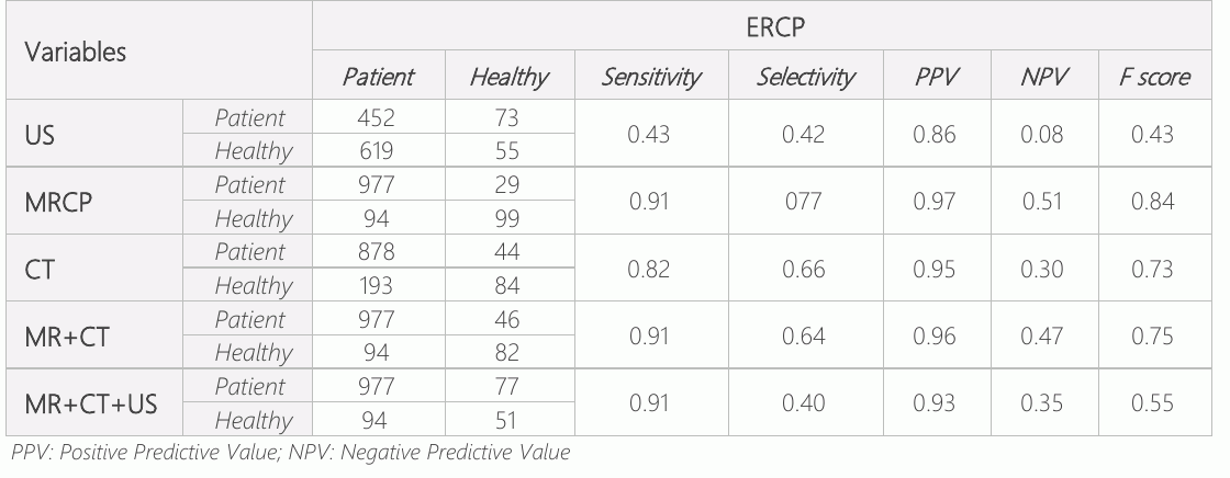
- Compared to ERCP, the most effective method in the diagnosis of choledocholithiasis is MRCP (F score: 0.84), CT (F score: 0.73), MR+BT (F score: 0.75), MR+BT+USG (F score: 0.55), and US (F score: 0.43).
- Conclusions
- ERCP is the gold standard in treating stones in the common bile duct. However, since this method is invasive, its use for diagnostic purposes has decreased due to the possibility of complications. In diagnosing common choledochal stones, MRCP alone is sufficient regardless of other imaging methods.
Keywords
Full text
Introduction
Choledocholithiasis remains one of the common medical problems leading to surgery. Endoscopic Retrograde Cholangiopancreatography (ERCP) is a method that can be used to diagnose and treat choledocholithiasis, but complications, presenting as pancreatitis, can occur in 8% to 12% (1, 2). Due to its invasiveness and complications, its use for diagnostic purpose has gradually decreased (3). However, ERCP should be preferred in patients with a strong suspicion of choledochal stones; otherwise, the use of ultrasonography (US), computed tomography (CT), or MR Cholangio pancreatography (MRCP) is preferred (4).
Abdominal US is a method used in the diagnosis of choledocholithiasis. However, Wilcox et al. (5) and Qiu et al. (6) reported two studies showing the presence of choledochal stones with different technique and percentage in patients with regular liver function tests and US findings. CT is preferred in emergencies to evaluate abdominal pain. The diagnostic value of tomography in this situation is controversial, and it is stated in the literature that it should not be preferred as a priority (7). MRCP and endoscopic USG are routinely preferred radiological methods today to avoid invasive procedures such as ERCP. Especially, MRCP is now recognized as the most appropriate non-invasive method to detect choledochal stones, with a sensitivity of 93% and a specificity of 96% (8).
As a result of obstructive jaundice due to choledochal stone, serious complications such as cholangitis, sepsis, multi-organ failure, coagulopathy, and kidney failure may develop (9). For this reason, the correct and early diagnosis of suspected cases of biliary tract obstruction is crucial in determining the appropriate treatment option (9). Some studies have shown the effectiveness of US, CT, and MRCP in the differential diagnosis of bile duct obstruction due to common choledochal stones (9). In this study, our objective was to determine the efficiency and superiority of US, CT, and MRCP in the diagnosis of patients who underwent ERCP with the preliminary diagnosis of choledocholithiasis.
Materials and Methods
Study Design
Between June 2019 and April 2022, a total of 1152 cases diagnosed with bile duct stones in Şanlıurfa Mehmet Akif İnan Training and Research Hospital General Surgery Clinic were included in the study.
Ethical Statement
The ethics committee approval of the study was obtained from Harran University Clinical Research Ethics Committee (HRÜ/22.15.03). All patients underwent contrast-enhanced abdominal CT at the time of admission to the clinic and US and MRCP of the entire abdomen 1-2 days after admission. Radiologists reported radiological examinations. Imaging studies revealed pancreaticobiliary ducts, and the localization and cause of the pathology were detected. ERCP was performed on these patients in an average of 2-7 days. F1 scores were calculated (10). The F1 score was taken as the basis for the evaluation of the results of this study (Table 1). The F1 score value shows the harmonic average of the sensitivity and specificity values (11).
Results
The mean age of 1152 patients who underwent ERCP for choledochal stone was 58.5±20.2 years. The gender distribution was 754 women (65.4%) and 398 (34.6%) men.
Discussion
In this study, the effectiveness of MRCP for detecting choledochal stones was restored. However, the diagnostic value of US, which is often used to evaluate common bile ducts, was deficient. In addition, the combined test (MR+CT and MR+CT+US) used in diagnosing choledochal stones did not increase the effectiveness.
Abdominal US is a noninvasive, inexpensive, practical examination that can be applied almost anywhere (3). Wilcox et al. (5) and Qiu et al. (6) reported two studies showing the presence of choledochal stones with different techniques and percentages in patients with routine liver function tests and in the US. That is why the abdominal US should be the first option, but if choledocholithiasis is suspected, it is necessary to perform further radiological examinations in addition to routine serology and US evaluation (7). In our study, the diagnostic value of the US was found to be low. While it was sufficient to detect patients with stones [positive predictive value (PPV): 0.86], it was very insufficient to exclude patients without stones [negative predictive value (NPV): 0.08]. In the study of Kalthantaner et al., the sensitivity of MRCP for choledocholithiasis was reported in a wide range between 0.50-1 and the specificity between 0.83-1 (12). The diagnostic value of MRCP is relatively high in biliary obstructions, especially choledocholithiasis, and invasive examinations of ERCP can reduce the diagnostic value (12). In another study, the sensitivity of MRCP for choledocholithiasis in patients confirmed by ERCP was 100%, the specificity was 86%, and the accuracy was 96% (13). In another study, the sensitivity of MRCP in determining lesions in the common bile ducts was 85%, specificity 75%, PPV 89.5%, NPV 66.7%, and accuracy 82% (14). MRCP sensitivity (66.6%), specificity (82.8%), PPV (86.9%), and NPV (59.1%) for choledocholithiasis were reported when patients whom ERCP could not determine were excluded. The sensitivity of MRCP has been shown to increase to 91.9% when stones of 5 mm and larger are taken into account (15). In our study, the sensitivity of MRCP was 0.91, selectivity 077, PPV 0.97, NPV0.51, and F score 0.84. These results are in agreement with previous studies.
There may be errors in the detection of choledochal stones in MRCP. Physiological changes, such as dysfunction of the sphincter of Oddi, which appear pathologically as stones or distal common bile duct tumors, and various technical artifacts, such as benign strictures, may occur. Due to this, it is known that small stones cannot be detected from time to time in MRCP (16, 17). Routine MRCP without positive laboratory tests for choledochal stones is the main limitation, as it is not affordable (18). Other drawbacks of MRCP are the possibility of being unable to be done in non-tertiary health institutions and the lack of therapeutic effect. Therefore, techniques that can be used for therapeutic and diagnostic use should be preferred in the suspicion of choledochal stones. Performing MRCP is also difficult in patients with severe obesity, having a metallic foreign body in their body, and claustrophobia. It is known that the presence of stones smaller than 5 mm and peripancreatic edema are disadvantageous in detecting choledochal stones (19). Despite many disadvantages, it is a superior technique to USG because it is non-invasive and provides a higher spatial and three-dimensional image with proprietary software (7). However, since it is a noninvasive technique, it should always be preferred before referring patients to other invasive procedures. CT is usually preferred in emergencies to evaluate abdominal pain. The diagnostic value of tomography in this situation is controversial, and it is stated in the literature that it should not be preferred as a priority (7). Because the lack of calcium in many gallstones leads to a similar density to surrounding bile, the CT's visibility and sensitivity are limited (7). CT scanning increases patients' exposure to X-rays and incurs higher costs than in the US (7). The diagnostic power of CT is higher than the US, and MRCP is lower in the diagnosis of common choledochal stones (20, 21). In some studies, CT cholangiography has similar sensitivity and specificity to MRCP (21, 22). The new multidetector CT showed a sensitivity of 78% and a specificity of 96% (23). On the other hand, Kim et al. (24) reported that in 17% of cases in 2013, 64-detector CT did not display choledochal stones due to <5 mm size, black/brown color, and age of the patient. Greater exposure to radiation and lower diagnostic power makes CT less effective than MRCP (7).
MRCP has been reported to be more sensitive than USG and CT in detecting common choledochal stones. MRCP has comparable sensitivity and specificity to ERCP in detecting extrahepatic bile duct stones and has been reported to be superior to ERCP in diagnosing intrahepatic bile duct stones (25). Studies have reported that the combination of MRCP and US-CT can show the degree of biliary obstruction with higher accuracy, thus enabling diagnosis in cases where the guidewire cannot advance due to closed obstruction and ERCP is not diagnostic (26). Although MRCP has high sensitivity and specificity, the sensitivity did not change positively when we performed the combined test application (MR-CT and MR-CT- US combinations) to increase sensitivity and selectivity. On the other hand, we observed that the selectivity gradually decreased and false positives increased with each new addition of the test, as expected in the parallel test application.
Conclusion
Although anamnesis, clinical, and laboratory findings often give an idea in diagnosing common choledochal stones, imaging in the early period is very important in making the diagnosis and avoiding unnecessary ERCP procedures. MRCP is a non-invasive, easily accessible method comparable to ERCP to differentiate choledocholithiasis. The fastest and most sensitive method should be determined to avoid unnecessary imaging. Choosing the imaging method according to clinical and laboratory findings is crucial for correct diagnosis.
Declarations
- Author contributions (CRediT)
- All authors contributed equally to all 14 CRediT roles (Conceptualization, Data curation, Formal analysis, Funding acquisition, Investigation, Methodology, Project administration, Resources, Software, Supervision, Validation, Visualization, Writing – original draft, Writing – review & editing).
- Funding
-
No financial support was provided from any institution or organization within the scope of the research.
- Conflicts of interest
- The authors declare no conflict of interest for the present study.
- Ethics approval
- The ethics committee approval of the study was obtained from Harran University Clinical Research Ethics Committee (HRÜ/22.15.03).
- Data availability
- A data availability statement was not reported in the published version of this article.
References (26)
20 of 26 references are linked to their DOI.
- Williams EJ, Taylor S, Fairclough P, Hamlyn A, Logan RF, Martin D, et al. Risk factors for complication following ERCP; results of a large -scale, prospective multicenter study. Endoscopy. 2007;39(9):793-801 DOI Crossref PubMed Google Scholar
- Freeman ML, Nelson DB, Sherman S, Haber GB, Herman ME, Dorsher PJ, et al. Complications of endoscopic biliary sphincterotomy. N Engl J Med. 1996;335(13):909-18 DOI Crossref PubMed Google Scholar
- Committee ASoP, Maple JT, Ben-Menachem T, Anderson MA, Appalaneni V, Banerjee S, et al. The role of endoscopy in the evaluation of suspected choledocholithiasis. Gastrointest Endosc. 2010;71(1):1-9 DOI Crossref PubMed Google Scholar
- Karakan T, Cindoruk M, Alagozlu H, Ergun M, Dumlu S, Unal S. EUS versus endoscopic retrograde cholangiography for patients with intermediate probability of bile duct stones: a prospective randomized trial. Gastrointest Endosc. 2009; 69(2):244-52 DOI Crossref PubMed Google Scholar
- Wilcox CM, Kim H, Trevino J, Ramesh J, Monkemuller K, Varadarajulu S. Prevalence of normal liver tests in patients with choledocholithiasis undergoing endoscopic retrograde cholangiopancreatography. Digestion. 2014;89(3):232-8 12 77 MRCP, CT and US in Choledochal Stones Ugurlu et al. DOI Crossref PubMed Google Scholar
- Qiu Y, Yang Z, Li Z, Zhang W, Xue D. Is preoperative MRCP necessary for patients with gallstones? An analysis of the factors related to missed diagnosis of choledocholithiasis by preoperative ultrasound. BMC Gastroenterol. 2015;15:158 DOI Crossref PubMed Google Scholar
- Cianci P, Restini E. Management of cholelithiasis with choledocholithiasis: Endoscopic and surgical approaches. World J Gastroenterol. 2021;27(28):4536-54 DOI Crossref PubMed Google Scholar
- Lammert F, Gurusamy K, Ko CW, Miquel JF, Mendez-Sanchez N, Portincasa P, et al. Gallstones. Nat Rev Dis Primers. 2016; 2:16024 PubMed Google Scholar
- Gutt C, Schlafer S, Lammert F. The Treatment of Gallstone Disease. Dtsch Arztebl Int. 2020;117(9):148-58 DOI Crossref PubMed Google Scholar
- Cohen JF, Korevaar DA, Altman DG, Bruns DE, Gatsonis CA, Hooft L, et al. STARD 2015 guidelines for reporting diagnostic accuracy studies: explanation and elaboration. BMJ Open. 2016;6(11):e012799 DOI Crossref PubMed Google Scholar
- Sasaki YJTtm. The truth of the F-measure. 2007;1(5):1-5 PubMed Google Scholar
- Kaltenthaler EC, Walters SJ, Chilcott J, Blakeborough A, Vergel YB, Thomas S. MRCP compared to diagnostic ERCP for diagnosis when biliary obstruction is suspected: a systematic review. BMC Med Imaging. 2006;6:9 DOI Crossref PubMed Google Scholar
- Gautier G, Pilleul F, Crombe-Ternamian A, Gruner L, Ponchon T, Barth X, et al. Contribution of magnetic resonance cholangiopancreatography to the management of patients with suspected common bile duct stones. Gastroenterol Clin Biol. 2004;28(2):129-34 DOI Crossref PubMed Google Scholar
- Bostanci E, Yol S, Ozogul Y, Kayaalp C, Atalay F, Akoglu MJTJoS. Comparison of magnetic resonance cholangio pancreatography with endoscopic retrograde cholangio pancreatography in patients with suspected common bile duct stone. 2003;19(3):157-62 PubMed Google Scholar
- Baykan AR, Buran T, Gerçeker E, Yüceyar H, Tarhan S, Kasap E, et al. Hepatobiliyer Hastalıklarda Tanıya Yönelik Görüntüleme Yöntemlerinden Endoskopik Retrograd Kolanjio pankreatografi ve Manyetik Rezonans Kolanjio pankreatografinin Etkinliğinin Karşılaştırılması.2021;8(1):116-21 DOI Crossref PubMed Google Scholar
- Dusunceli E, Erden A, Erden I. [Anatomic variations of the bile ducts: MRCP findings]. Tani Girisim Radyol. 2004;10(4):296- PubMed Google Scholar
- Srinivasa S, Sammour T, McEntee B, Davis N. Selective use of magnetic resonance cholangiopancreatography in clinical practice may miss choledocholithiasis in gallstone pancreatitis. Can J Surg. 2010;53(6):403-7 PubMed Google Scholar
- Epelboym I, Winner M, Allendorf JD. MRCP is not a cost-effective strategy in the management of silent common bile duct stones. J Gastrointest Surg. 2013;17(5):863-71 DOI Crossref PubMed Google Scholar
- Polistina FA, Frego M, Bisello M, Manzi E, Vardanega A, Perin B. Accuracy of magnetic resonance cholangiography compared to operative endoscopy in detecting biliary stones, a single center experience and review of literature. World J Radiol. 2015;7(4):70-8 DOI Crossref PubMed Google Scholar
- Mitchell SE, Clark RA. A comparison of computed tomography and sonography in choledocholithiasis. AJR Am J Roentgenol. 1984;142(4):729-33 DOI Crossref PubMed Google Scholar
- Soto JA, Alvarez O, Munera F, Velez SM, Valencia J, Ramirez N. Diagnosing bile duct stones: comparison of unenhanced helical CT, oral contrast-enhanced CT cholangiography, and MR cholangiography. AJR Am J Roentgenol. 2000;175(4): 1127-34 DOI Crossref PubMed Google Scholar
- Kondo S, Isayama H, Akahane M, Toda N, Sasahira N, Nakai Y, et al. Detection of common bile duct stones: comparison between endoscopic ultrasonography, magnetic resonance cholangiography, and helical-computed-tomographic cholangiography. Eur J Radiol. 2005;54(2):271-5 DOI Crossref PubMed Google Scholar
- Anderson SW, Rho E, Soto JA. Detection of biliary duct narrowing and choledocholithiasis: accuracy of portal venous phase multidetector CT. Radiology. 2008;247(2):418-27 DOI Crossref PubMed Google Scholar
- Kim CW, Chang JH, Lim YS, Kim TH, Lee IS, Han SW. Common bile duct stones on multidetector computed tomography: attenuation patterns and detectability. World J Gastroenterol. 2013;19(11):1788-96 DOI Crossref PubMed Google Scholar
- Kim TK, Kim BS, Kim JH, Ha HK, Kim PN, Kim AY, et al. Diagnosis of intrahepatic stones: superiority of MR cholangiopancreatography over endoscopic retrograde cholangiopancreatography. AJR Am J Roentgenol. 2002; 179(2):429-34 DOI Crossref PubMed Google Scholar
- Hacim NA, Akbas A, Meric S, Karabay O, Ulgen Y, Tokocin M, et al. Predictive value of ultrasonography and magnetic resonance cholangiopancreatography in the diagnosis of biliary obstruction. Ann Ital Chir. 2020;91:277-82 PubMed Google Scholar
Respond to this article
Comments on a published article are welcome as a Letter to the Editor.
Get an e-mail when new articles in your fields are published
Questions to the authors
Questions are moderated: an editor approves each question, the authors answer, and an editor approves the answer before both appear here.
No answered questions yet.
Ask the authors a question
Discussion
Comments are moderated: they appear after the writer confirms the e-mail address and an editor approves them. Each comment carries a conflict-of-interest statement.
No comments yet.
Write a comment
Read the full text
Open the PDF here
Loading the PDF…
If the PDF does not show here, use the download button above.
How to cite this article
Ugurlu ET, Dere O, Yilmaz S. Comparison of the Diagnostic Value of MRCP, CT, and Ultrasonography in Choledochal Stones with the Results of ERCP: Evidence from 1152 Case. Ulutas Med J. 2023;9(1):8-12. doi:10.5455/umj.20221205103820
Ugurlu, E. T., Dere, O., & Yilmaz, S. (2023). Comparison of the Diagnostic Value of MRCP, CT, and Ultrasonography in Choledochal Stones with the Results of ERCP: Evidence from 1152 Case. The Ulutas Medical Journal, 9(1), 8–12. https://doi.org/10.5455/umj.20221205103820
Ugurlu, E.T., Dere, O. and Yilmaz, S. (2023) 'Comparison of the Diagnostic Value of MRCP, CT, and Ultrasonography in Choledochal Stones with the Results of ERCP: Evidence from 1152 Case', The Ulutas Medical Journal, 9(1), pp. 8–12. doi: 10.5455/umj.20221205103820.
@article{umj80098,
  title = {Comparison of the Diagnostic Value of MRCP, CT, and Ultrasonography in Choledochal Stones with the Results of ERCP: Evidence from 1152 Case},
  author = {Ugurlu, Esat Taylan and Dere, Osman and Yilmaz, Sezgin},
  journal = {The Ulutas Medical Journal},
  year = {2023},
  volume = {9},
  number = {1},
  pages = {8--12},
  doi = {10.5455/umj.20221205103820},
  url = {https://www.ulutasmedicaljournal.com/?mno=80098},
  issn = {2149-388X}
}