The Ulutas Medical Journal · ISSN 2149-0430 (print), 2149-388X (online) · https://www.ulutasmedicaljournal.com/?mno=87160
Case Report · Open access
Intrabiliary Metastasis of Colon Cancer Mimicking Cholangiocarcinoma and Chemotherapy-Induced Sclerosing Cholangitis: A Case Report
Also available in: العربية · Türkçe · 中文
- Department of Radiology, Chi Mei Medical Center, Liouying, Tainan, Taiwan ROR
- Department of Intensive Care Medicine, Chi Mei Medical Center, Liouying, Tainan, Taiwan ROR
- Department of Nursing, Kaohsiung Municipal United Hospital, Taiwan ROR
Abstract
Intrabiliary metastasis of colon cancer without the involvement of the hepatic parenchyma is very rare and can cause biliary obstruction. We herein present a rare case of colon cancer metastasis that developed after right hemicolectomy and while undergoing chemotherapy. Computed tomography (CT) of the abdomen and percutaneous transhepatic cholangiography and drainage (PTCD) was performed because of obstructive jaundice. CT-guided biopsy showed high-grade atypical glands that were CK7(â'), CK20(+), and CDX-2(+); metastatic adenocarcinoma would be considered first rather than cholangiocarcinoma. Clinicians should be aware that intrabiliary metastasis of colon cancer can mimic cholangiocarcinoma and chemotherapy-induced sclerosing cholangitis.
Keywords
Full text
Introduction
Intrabiliary metastasis of colon cancer without involvement of the hepatic parenchyma is rare (1). Metastases to the liver can cause biliary obstruction and result in painless jaundice, pale stools, dark urine, and pruritis, and such metastases are associated with elevations of direct bilirubin and alkaline phosphatase (2). In addition, imaging findings showing such metastases as mimicking sclerosing cholangitis as a multifocal biliary stricture are rare. Here, we report a case of intrabiliary metastasis of colon cancer and the associated imaging findings.
Case Presentation
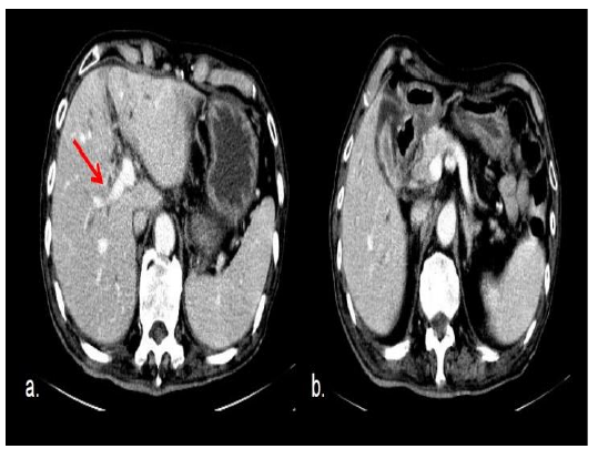
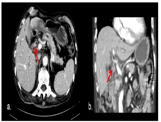
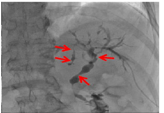
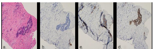
An 82-year-old man presented with colon metastasis after right hemicolectomy and while undergoing chemotherapy. Laboratory tests at regular follow-up revealed elevated CA19-9 and CEA. Computed tomography (CT) was performed, which revealed soft tissue density in the common bile duct (CBD) and right intrahepatic duct (IHD) with wall thickening, resulting in biliary dilatation (Figures 1 and 2). Percutaneous transhepatic cholangiography and drainage (PTCD) was performed, and cholangiography revealed multiple strictures of the intrahepatic and extrahepatic bile ducts, causing obstructive jaundice (Figure 3). Based on his history and radiological findings, the differential diagnosis included metastasis, chemotherapy-induced sclerosing cholangitis (CISC), and cholangiocarcinoma. Cytology of bile collected from the percutaneous transhepatic drainage tube revealed no features of malignancy. CT-guided biopsy was then performed and revealed high-grade atypical glands. Immunohistochemical study revealed that the atypical glands were CK7(−), CK20(+), and CDX-2(+) (Figure 4).
According to the study result, metastatic adenocarcinoma would be considered first rather than biliary duct primary malignancy given the absence of CK7 expression. The patient’s condition was poor, total bilirubin levels increased again, and liver function worsened, requiring the insertion of two PTCDs on each side. One month later, septic biliary tract infection exacerbated progressively, and the patient finally died.
Discussion
In our case, CT and cholangiography revealed wall thickening of the extrahepatic and intrahepatic bile ducts accompanied by stricture and dilatation. Our case also presented the imaging findings of biliary obstruction and biliary stricture, which are also the imaging findings for secondary sclerosing cholangitis and primary cholangiocarcinoma. CISC has been reported to be occasionally caused by hepatic arterial infusion chemotherapy with fluoropyrimidines (3–5). In the study, considering the sudden onset of bile duct lesion during chemotherapy (FOLFOX4), as sclerosing cholangitis was suspected, the patient received initial treatment with steroids, and the chemotherapy was discontinued. However, rapid elevated tumor markers (CEA: 171 ng/mL, CA19-9: 10115 U/mL) and no improvement after cessation of chemotherapy were noted. Distinctive findings of sclerosing cholangitis, such as “onion skin” fibrosis (6) around the bile duct, were not found in the liver biopsy in our case. Cholangiocarcinoma has the following morphology and growth patterns: mass formation, periductal infiltration, and intraductal growth (7). The imaging findings of intraductal and periductalinfiltrating type of cholangiocarcinoma included biliary strictures, intraductal neoplasms, and focal or diffuse ductal thickening with or without enhancement (8, 9). Differentiating intrabiliary colorectal liver metastases from primary intrahepatic cholangiocarcinoma is difficult. However, immunohistochemical studies, such as CK7, CK20, and CDX2 staining, can provide the definite diagnosis (10). Metastatic colon carcinoma shows CK20 positivity and CK7 negativity in contrast to the adjacent bile duct epithelium, which is CK7-positive and CK20- negative (11). In our case, no CK7 expression was found. According to the result, metastatic adenocarcinoma would be considered first rather than primary biliary duct malignancy. In our case, imaging studies revealed obstructive jaundice without an intraparenchymal mass. Biopsy techniques for intrabiliary lesions include brush cytology, clamshell forceps under choledochoscopic guidance, and clamshell forceps under fluoroscopic guidance. In a previous study, the choledochoscope-directed biopsy technique had highest sensitivity and specificity, but this difference was not statistically significant (12). However, in this study, the patient refused to accept endoscopic retrograde cholangiopancreatography (ERCP), and clamshell forceps were not available in our hospital; thus, we performed transhepatic biopsy. Because ductal thickening with soft tissue density cannot be well identified under sonography, real-time USG combined with CT is useful for localizing inconspicuous focal hepatic lesions (13). The etiology of intrabiliary growth of liver metastasis is unknown, but the etiology probably involves vascular invasion or the implantation of malignant cells in the biliary epithelium through bile flow (14). Patients with intrabiliary metastasis have better prognosis than patients without metastasis; moreover, patients with macroscopic involvement have a much higher 5-year survival rate (80%) than those with microscopic involvement (15). However, the condition of our patient was poor, his total bilirubin levels increased again, and his liver function worsened, even after two PTCDs were inserted on both sides; consequently, the patient was transferred to the hospice ward and died 1 month later.
Conclusion
In conclusion, intrabiliary metastasis is easily overlooked. An immunohistochemical study is helpful to achieve the correct diagnosis and management.
Declarations
- Author contributions (CRediT)
- All authors contributed equally to all 14 CRediT roles (Conceptualization, Data curation, Formal analysis, Funding acquisition, Investigation, Methodology, Project administration, Resources, Software, Supervision, Validation, Visualization, Writing – original draft, Writing – review & editing).
- Funding
-
Funding information was not reported in the published version of this article.
- Conflicts of interest
- All authors declare that there is no conflict of interest.
- Ethics approval
- Ethics approval details were not reported in the published version of this article.
- Data availability
- A data availability statement was not reported in the published version of this article.
References (15)
14 of 15 references are linked to their DOI.
- Patel T. Increasing incidence and mortality of primary intrahepatic cholangiocarcinoma in the United States. Hepatology. 2001;33:1353–1357 DOI Crossref PubMed Google Scholar
- Nagler J, Rochwarger AM. Metastatic colon carcinoma simulating primary bile duct carcinoma via endoscopic cholangiography. Gastrointest Radiol. 1977;2:75–76 DOI Crossref PubMed Google Scholar
- Sandrasegaran K, Alazmi WM, Tann M, et al. Chemo therapy-induced sclerosing cholangitis. Clin Radiol. 2006;61:670–8 DOI Crossref PubMed Google Scholar
- Shea WJ Jr, Demas BE, Goldberg HI, et al. Sclerosing cholangitis associated with hepatic arterial FUDR chemotherapy: Radiographic-histologic correlation. AJR Am J Roentgenol. 1986; 146:717–21 DOI Crossref PubMed Google Scholar
- Kemeny N, Daly J, Oderman P, et al. Hepatic artery pump infusion: Toxicity and results in patients with metastatic colorectal carcinoma. J Clin Oncol. 1984; 2:595-600 85 77 Intrabiliary Metastasis of Colon Cancer Guang-Mao Lee et al. DOI Crossref PubMed Google Scholar
- Akira K, Kazuyoshi O, Nobuyasu F, et al. Chemo therapy-Induced Sclerosing Cholangitis Caused by Systemic Chemotherapy, ACG Case Rep J 2019;6:1-4 PubMed Google Scholar
- Yamasaki S. Intrahepatic cholangiocarcinoma: macro scopic type and stage classification. J Hepato biliary Pancreat Surg 2003; 10:288-291 DOI Crossref PubMed Google Scholar
- Lee WJ, Lim HK, Jang KM, et al. Radiologic spectrum of cholangiocarcinoma: emphasis on manifestations and differential diagnoses. Radio Graphics 2001;21(spec issue):S97–S116 DOI Crossref PubMed Google Scholar
- Han JK, Choi BI, Kim AY, et al. Cholangiocarcinoma: pictorial essay of CT and cholangiographic findings. RadioGraphics 2002;22:173–187 DOI Crossref PubMed Google Scholar
- Dong Y, Patel H, Patel C. Intrabiliary hepatic metastasis of colorectal carcinoma mimicking primary cholangio carcinoma: a case report and review of the literature. Case Rep Pathol. 2016; 4704781 DOI Crossref PubMed Google Scholar
- Tokai H, Kawashita Y, Eguchi S, et al. A case of mucin producing liver metastases with intrabiliary extension. World J Gastroenterol 2006; 12:4918–4921 DOI Crossref PubMed Google Scholar
- Scott J. Savader, Carol A. Prescott, Gunnar B. Lund, et al. Intraductal biliary biopsy: comparison of three techniques. J Vasc Interv Radiol 1996;7(5):743-50 DOI Crossref PubMed Google Scholar
- Min Woo Lee. Fusion imaging of real-time ultrasono graphy with CT or MRI for hepatic intervention. Ultrasonography. 2014 Oct; 33(4): 227–239 DOI Crossref PubMed Google Scholar
- Kim AY, Jeong WK. Intraductal malignant tumors in the liver mimicking cholangiocarcinoma: imaging features for differential diagnosis. Clin Mol Hepatol. 2016;22: 192–7 DOI Crossref PubMed Google Scholar
- Raquel A. Latorre Fragua, Alba Manuel Vazquez, Yuri Rodrigues Figueira, et al. Intrabiliary metastases in colorectal cancer: a systematic review. J Hepatobiliary Pancreat Sci (2019) 26:270–280 DOI Crossref PubMed Google Scholar
Respond to this article
Comments on a published article are welcome as a Letter to the Editor.
Get an e-mail when new articles in your fields are published
Questions to the authors
Questions are moderated: an editor approves each question, the authors answer, and an editor approves the answer before both appear here.
No answered questions yet.
Ask the authors a question
Discussion
Comments are moderated: they appear after the writer confirms the e-mail address and an editor approves them. Each comment carries a conflict-of-interest statement.
No comments yet.
Write a comment
Read the full text
Open the PDF here
Loading the PDF…
If the PDF does not show here, use the download button above.
How to cite this article
Chen PY, Chao CM, Lee GM. Intrabiliary Metastasis of Colon Cancer Mimicking Cholangiocarcinoma and Chemotherapy-Induced Sclerosing Cholangitis: A Case Report. Ulutas Med J. 2022;8(2):82-85. doi:10.5455/umj.20220222072137
Chen, P. Y., Chao, C. M., & Lee, G. M. (2022). Intrabiliary Metastasis of Colon Cancer Mimicking Cholangiocarcinoma and Chemotherapy-Induced Sclerosing Cholangitis: A Case Report. The Ulutas Medical Journal, 8(2), 82–85. https://doi.org/10.5455/umj.20220222072137
Chen, P.Y., Chao, C.M. and Lee, G.M. (2022) 'Intrabiliary Metastasis of Colon Cancer Mimicking Cholangiocarcinoma and Chemotherapy-Induced Sclerosing Cholangitis: A Case Report', The Ulutas Medical Journal, 8(2), pp. 82–85. doi: 10.5455/umj.20220222072137.
@article{umj87160,
  title = {Intrabiliary Metastasis of Colon Cancer Mimicking Cholangiocarcinoma and Chemotherapy-Induced Sclerosing Cholangitis: A Case Report},
  author = {Chen, Po-Yuan and Chao, Chien-Ming and Lee, Guang-Mao},
  journal = {The Ulutas Medical Journal},
  year = {2022},
  volume = {8},
  number = {2},
  pages = {82--85},
  doi = {10.5455/umj.20220222072137},
  url = {https://www.ulutasmedicaljournal.com/?mno=87160},
  issn = {2149-388X}
}