The Ulutas Medical Journal · ISSN 2149-0430 (print), 2149-388X (online) · https://www.ulutasmedicaljournal.com/?mno=113681
Case Report · Open access
Melanonychia Striata: A Case Report
Also available in: العربية · Türkçe · 中文
- Surgical Oncology, Surgery, Udayana University, Sanglah General Hospital, Prima Medika Hospital, Denpasar, Bali, Indonesia ROR
- Faculty of Medicine, Udayana University, Sanjiwani General Hospital, Gianyar, Bali, Indonesia ROR
- Pathology, Udayana University, Sanglah General Hospital RS Prima Medika, Prima Medika Hospital, Denpasar, Bali, Indonesia ROR
Abstract
Subungual melanoma has higher mortality than other skin melanomas caused by a late initial diagnosis of this disease, leading to a poor prognosis. Two-thirds of subungual melanoma symptoms begin with melanonychia striata, appearing as brown or black-pigmented lines of the nail plate. In this case report, we presented a case of melanonychia striata in a 30-year-old male. Longitudinal excision or partial avulsion was performed, followed by histopathology examination to exclude malignancy and determine further management for this case.
Keywords
Full text
Introduction
The case of melanonychia striata (longitudinal melanonychia) is a condition in which the nail plate forms one or several brown or black-pigmented lines. This condition is caused by increased melanin production in the nail matrix, which is then stored in the nail plate. Proliferation and hyperplasia of melanocyte in the nail matrix can cause increased melanin production, besides bleeding or trauma (1). In Caucasian, the prevalence of melanonychia striata is 1.4%, most frequently occurred in the thumb, toes, and index fingers. While in Asian, the prevalence was found to be higher, reported as much as 20%-23% and 11.4% in the year 1933 and 1958, respectively (2).
Histopathology examination is needed since it is crucial to rule out the possibility of malignancy or melanoma in the case of melanonychia striata. In this case report, we presented a case of melanonychia striata in a 30-year-old male.
Case Presentation
A 30-year-old man was referred to the Surgical Oncology Clinic of Prima Medika Hospital with initial suspicion of subungual melanoma. The patient had discoloration of the first toenail of the left foot since six months ago (Figure 1). His nail was turning brownish-black, being darker over time, but painless. The patient was in good health and not taking any medication. There was no history of trauma and no similar complaints in his family. The patient also said that there was no family history of melanonychia striata, melanoma (or other diseases of the nail), other skin diseases such as psoriasis, atopic dermatitis, and others.
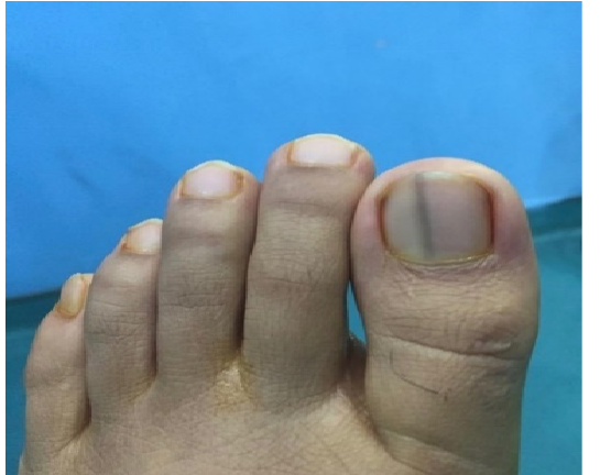
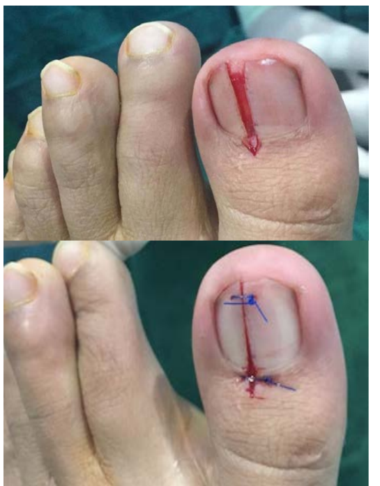
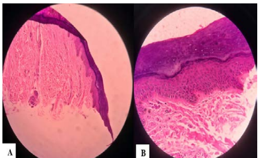
We found a longitudinal brownish-black discoloration on first left toenail, 1 mm width, on physical examination of this patient. The nail was neither fragile nor split. His other nails were normal. There was no skin discoloration around the nail. Diagnosis of melanonychia striata (longitudinal melanonychia) was made based on the patient's age, physical examination, and histopathology results. The specimen was obtained from longitudinal excision (partial nail avulsion) of the nail plate and matrix, stained with hematoxylin-eosin. Histopathology results showed that there was no proliferation of melanocytes or malignant melanoma. In this case, longitudinal excision (partial nail avulsion) of the nail with melanonychia striata was done for diagnostic procedures and therapeutic management (Figure 2).
Discussion
Melanonychia striata is a condition where the nail plate forms one or several brown or black-pigmented lines (1). Increased nail pigmentation is caused by the activation of nail matrix melanocytes, which causes an increase in pigment production. Mechanism of activation of nail melanocytes is not fully understood but is believed to be caused by excessive expression of the α-melanocyte-stimulating hormone, adrenocorticotropic hormone, and ultraviolet. Melanonychia usually shows benign lesions on the nail matrix related to melanocytic nevus, lentigo, or increased benign melanocyte activity. However, a melanonychia can also represent pathological conditions such as bacterial or fungal infection, melanoma, and other malignancies (3).
About two-thirds of subungual melanoma cases appear with melanonychia striata as an early symptom. Some approaches can be taken to exclude malignancy i.e., "wait and see" and longitudinal excision (which is chosen) (4, 5). In malignant case, other nail surgery such as amputation or wide excision usually is performed, with the aim of removing local tumors on the nail, treating nail infections, reducing pain due to trauma, making the diagnosis by biopsy, or aesthetic purpose (6). Nail plate surgery (nail avulsion) can be done either totally or partially. In this case, we performed partial nail avulsion for our patients. Compared with total nail avulsion, partial nail avulsion is preferred to reduce complications (bleeding, infection, pain in changing wound dressing, or during activities). Then, the main concern is to rule out the possibility of malignancy. In this patient, the histopathologic examination of longitudinal excision did not show any melanocyte proliferation (Figure 3). Discoloration of the nail might have resulted from physiologically increased a melanocyte function rather than pathologically hyperplasia due to melanocytic nevus, or even malignant melanoma. In our case, no malignancy sign was found. There were no hyperpigmentation more than 6 mm and nail dystrophy (5). However, in some cases, a change from benign to the malignant lesion can still occur. Therefore, it is important to perform monitoring examinations annually (6).
Conclusion
Melanonychia striata is a change of nail pigment, recognized mostly as an early sign of a subungual melanoma. Histopathological examination after excision must be done to exclude the possibility of malignancy.
Declarations
- Author contributions (CRediT)
- All authors contributed equally to all 14 CRediT roles (Conceptualization, Data curation, Formal analysis, Funding acquisition, Investigation, Methodology, Project administration, Resources, Software, Supervision, Validation, Visualization, Writing – original draft, Writing – review & editing).
- Funding
-
Funding information was not reported in the published version of this article.
- Conflicts of interest
- The authors declared no conflict of interest
- Ethics approval
- The Ethical Committee and Institutional Review Board where the present study was conducted, approved the study design. The patient has been consented that this case report will be submitted for publication.
- Data availability
- A data availability statement was not reported in the published version of this article.
References (6)
6 of 6 references are linked to their DOI.
- Lee M, Seo S, Jung J, Shin Y, Cho E, Park E, Kim K, Kim K. Longitudinal melanonychia in childhood: a clinical and histopathological review of Korean patients. European Journal of Dermatology. 2017;27(3):275-280 DOI Crossref PubMed Google Scholar
- Koga H, Saida T, Uhara H. Key point in dermoscopic differentiation between early nail apparatus melanoma and benign longitudinal melanonychia. The Journal of Derma tology. 2010:38(1):45-52 DOI Crossref PubMed Google Scholar
- Ehsanzadeh-Cheemeh P, Grimes R, Rowan P, Huang Y, Essien E, Lewis S. Melanonychia in Patients Infected with Human Immunodeficiency Virus Original Communication. Advances in Infectious Diseases. 2011;1(2): 15-19 DOI Crossref PubMed Google Scholar
- Ruben BS, Pigmented lesions of the nail unit: clinical and histopathologic features, Semin Cutan Med Surg. 2010; 29(3):148-158 DOI Crossref PubMed Google Scholar
- Skornšek N, Orešič Barač T, Marko P. Congenital longitudinal melanonychia: a case report. Acta Dermatovenerologica Alpina Pannonica et Adriatica. 2017;26(4);55-59 DOI Crossref PubMed Google Scholar
- Saraswati N, Sutedja E, Agusni J. Berbagai Prosedur Bedah Kuku. Syifa' MEDIKA: Jurnal Kedokteran dan Kesehatan. 2019; 8(1):15 Melanonychia Striata Adiputra et al. DOI Crossref PubMed Google Scholar
Respond to this article
Comments on a published article are welcome as a Letter to the Editor.
Get an e-mail when new articles in your fields are published
Questions to the authors
Questions are moderated: an editor approves each question, the authors answer, and an editor approves the answer before both appear here.
No answered questions yet.
Ask the authors a question
Discussion
Comments are moderated: they appear after the writer confirms the e-mail address and an editor approves them. Each comment carries a conflict-of-interest statement.
No comments yet.
Write a comment
Read the full text
Open the PDF here
Loading the PDF…
If the PDF does not show here, use the download button above.
How to cite this article
Adiputra PAT, Alviariza A, Saputra H. Melanonychia Striata: A Case Report. Ulutas Med J. 2020;6(3):176-179. doi:10.5455/umj.20200728095745
Adiputra, P. A. T., Alviariza, A., & Saputra, H. (2020). Melanonychia Striata: A Case Report. The Ulutas Medical Journal, 6(3), 176–179. https://doi.org/10.5455/umj.20200728095745
Adiputra, P.A.T., Alviariza, A. and Saputra, H. (2020) 'Melanonychia Striata: A Case Report', The Ulutas Medical Journal, 6(3), pp. 176–179. doi: 10.5455/umj.20200728095745.
@article{umj113681,
  title = {Melanonychia Striata: A Case Report},
  author = {Adiputra, Putu Anda Tusta and Alviariza, Annisa and Saputra, Herman},
  journal = {The Ulutas Medical Journal},
  year = {2020},
  volume = {6},
  number = {3},
  pages = {176--179},
  doi = {10.5455/umj.20200728095745},
  url = {https://www.ulutasmedicaljournal.com/?mno=113681},
  issn = {2149-388X}
}