The Ulutas Medical Journal · ISSN 2149-0430 (print), 2149-388X (online) · https://www.ulutasmedicaljournal.com/?mno=25913
Case Report · Open access
Arrested Pneumatization of the Sphenoid Sinus: A Normal Variant That Can Be Confused With a Pathological Entity
- Cumhuriyet University, School of Medicine, Department of Radiology, Sivas, Turkey ROR
Abstract
- Background
- An arrested pneumatization of the sphenoid sinus is an anatomical variant. It is known that the sphenoid bones undergo early fatty marrow conversion antecedent to normal pneumatization. Since it is a developmental condition, it usually occurs as a coincidental finding. The clinical symptoms specific to this entity are not attributable. They are seen incidentally during radiological examinations. If not recognized, it can be easily confused with pathological lesions. In this article, we aimed to draw attention to this rare situation by observing the current literature with three cases.
- Conclusion
- In conclusion, making a correct diagnosis of AP of sphenoid sinus, an incidentally detected benign, asymptomatic developmental variation, and making a careful distinction of it from other lesions will save patients from unnecessary biopsy and surgical procedures.
Keywords
Full text
Introduction
A pause in the customarily expected pneumatization of the sphenoid sinus is termed as arrested pneumatization (AP) of sphenoid sinus (incomplete pneumatization, sphenoid pseudolesion). It is an incidental radiological sign on computed tomography and magnetic resonance imaging, which can be confused with aggressive pathological conditions. Although AP is a benign, asymptomatic condition, it is imperative to make its radiological diagnosis to avoid unnecessary invasive interventions (1–5). AP of the sphenoid sinus is rarely seen in the literature. Recently, an increase in similar cases is noticed. However, in search of Turkish literature, we did not find any publication on the subject. In this article, we aimed to draw attention to this rare situation by observing the current literature with three cases.
Case presentation
Case 1
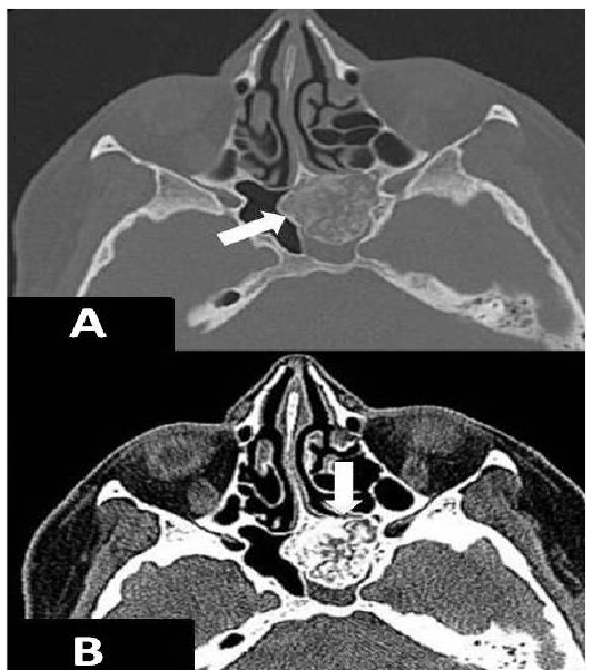
A temporal bone computed tomography (CT) examination for chronic otitis media in 51-year-old woman revealed a non-expansile lesion of soft tissue density located intrasinusal in the left half of the sphenoid sinus, which contained a sclerotic margin, curvilinear calcifications, and patchy areas of fat density (Figures 1A and 1B). No mass effect or destruction was evident in the adjacent bony structures.
Case 2
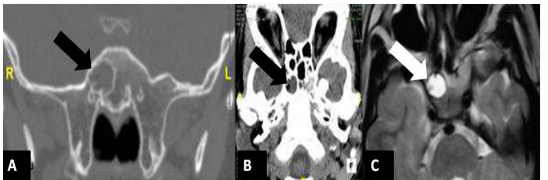
A cranial magnetic resonance imaging (MRI) examination for headache in a 7-year-old boy revealed a hyperintense, non-expansile lesion on T1 and T2 weighted series inside the sphenoid sinus on the right side. A CT examination of this region showed a non-expansile, non-calcified lesion of soft tissue density in the right half of the sphenoid sinus. The left side of the sphenoid sinus did not show aeration (Figures 2A-C). No mass effect or destructive changes were present in bones adjacent to the described lesion.
Case 3
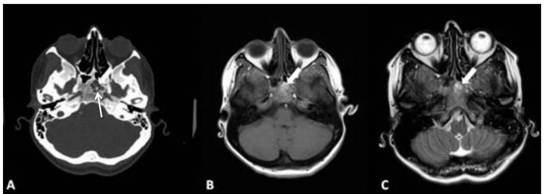
A cranial MRI examination of a 38-year-old woman with headache, intermittent nausea, and vomiting revealed a lesion with the hyperintense signal on T1 and T2 weighted series showing no mass effect or destructive properties in the left half of the sphenoid sinus. A CT examination showed that the lesion had a sclerotic margin and a central calcified area (Figures 3A-C). Physical and neurological examination and laboratory tests of the patients were unremarkable. Further studies did not unearth any hematological disorder. The described lesions of the patients were diagnosed as AP based on CT and MRI findings.
Discussion
Paranasal sinus pneumatization is a long-term developmental process. It starts in utero and continues into adulthood. Pneumatization is quite variable, both developmentally and anatomically. Arrested pneumatization mostly develops in the sphenoid sinus but may also involve maxillary and frontal sinuses. The absence of otherwise expected pneumatization in bones forming the base of the head is termed as AP. AP is a developmental variation that most commonly involves the sphenoid sinus. It is known that conversion in the fatty bone marrow of the sphenoid bone is the precursor of pneumatization. Conversion from red marrow to fatty marrow in the sphenoid sinus starts at the presphenoidal region anteriorly and proceeds to clivus posteriorly. This process starts at the seventh postnatal month. However, the actual conversion is more common at around age 2. This anterior-posterior bone marrow conversion is closely related to sphenoid sinus pneumatization (1–6). However, pneumatization of the sphenoid sinus is a dynamic process. Around 14 years, the expansion is completed, but pneumatization progresses slowly and is wholly discontinued until the age of 25 years. (7).
In individuals with AP, the processes of pneumatization of the sphenoid sinus and its lining with respiratory epithelium are arrested as a result of the normally expected conversion of red marrow to fatty marrow and the following involution of fat tissue. However, certain factors such as ambient temperature, bone composition, and/or vascularization are also thought to play a role in this arrest. Prabhu and Branstetter revealed that the prevalence of AP in patients with sickle cell anemia is increased compared to healthy people owing to regional blood flow anomalies or increased serum erythropoietin level (5). If developmental variation and its differential diagnoses are not adequately known, the difficulty arises in the interpretation of CT and MRI examinations, and unnecessary operations may ensue. AP is diagnosed incidentally when it is asymptomatic. No specific symptoms have been attributed to this condition. It generally involves regions related to the actual or accessory sphenoid sinus, such as the floor of the sphenoid sinus, pterygoid process, and clivus (1–3, 6, 8).
The best markers of the non-expansile character of AP on CT and MRI of the base of the skull include the absence of displacement or morphological disruption in the Vidian canal and inferior orbital sinus. In particular, because the Vidian nerve is located just below the sphenoid sinus mucosa, an unnecessary invasive procedure for this region can cause damage to the Vidian nerve (9). It appears as a non-expansile lesion that contains curvilinear calcification and a sclerotic margin on CT. On MRI, internal fat and microcysts that do not exert a mass effect are diagnostic. This internal fat content is hyperintense, whereas microcysts, calcified areas, and sclerotic appear hypointense on T1-weighted MRI images. On T2-weighted imaging microcystic component is hyperintense. MRI series with gadolinium enhancement show no contrast uptake. While CT is more effective in showing the osseous components of the lesions, MRI indicates the internal structure of the lesions and contributes to the differential diagnosis (6–8, 11, 12).
The radiological criteria suggesting an AP of the central base of the head were defined by Welker et al. (8):
• An incidental finding on CT imaging, • Asymptomatic abnormal ossification zone, • Presence of the central base of the head in a place where usual pneumatization is found. Differential diagnosis of AP includes fibrous dysplasia, intraosseous lipoma, intraosseous hemangioma, ossifying fibroma, chondro sarcoma, skull base osteomyelitis, chondroid chordoma, and bone metastases. Unlike AP, however, all these pathological conditions display signs of mass effect in the form of a loss of internal fat content or compression, displacement, or destruction of adjacent structures (2–6, 8, 13). The conchal type of sphenoid sinus pneumatization should also be considered in the differential diagnosis of AP. The conchal type of sphenoid sinus pneumatization is more common in children, and it is rare in adults (7).
In conclusion, making a correct diagnosis of AP of sphenoid sinus, an incidentally detected benign, asymptomatic developmental variation, and making a careful distinction of it from other lesions will save patients from unnecessary biopsy and surgical procedures.
Declarations
- Author contributions (CRediT)
- All authors contributed equally to all 14 CRediT roles (Conceptualization, Data curation, Formal analysis, Funding acquisition, Investigation, Methodology, Project administration, Resources, Software, Supervision, Validation, Visualization, Writing – original draft, Writing – review & editing).
- Funding
-
Funding information was not reported in the published version of this article.
- Conflicts of interest
- The author declares no conflict of interests.
- Ethics approval
- Ethics approval details were not reported in the published version of this article.
- Data availability
- A data availability statement was not reported in the published version of this article.
References (13)
11 of 13 references are linked to their DOI.
- Kuntzler S, Jankowski R. Arrested pneumatization: the witness of paranasal sinuses development? Eur Ann Otorhinolaryngol Head Neck Dis. 2014; 131: 167-70 DOI Crossref PubMed Google Scholar
- Jalali E, Tadinada A. Arrested pneumatization of the sphenoid sinus mimicking intraosseous lesions of the skull base. Imaging Sci Dent. 2015; 45: 67-72 DOI Crossref PubMed Google Scholar
- De Jong MD, Fassaert TA, Ranschaert ER. Arrested pneumatization of the skull base JBR-BTR. 2011;94:114-5 DOI Crossref PubMed Google Scholar
- Aoki S, Dillon WP, Barkovich AJ, Norman D. Marrow conversion before pneumatization of the sphenoid sinus: assessment with MR imaging. Radiology 1989; 172:373-5 DOI Crossref PubMed Google Scholar
- Prabhu AV, Branstetter BF IV. The CT Prevalence of Arrested Pneumatization of the Sphenoid Sinus in Patients with Sickle Cell Disease. AJNR Am J Neuroradiol. 2016; May 5 DOI Crossref PubMed Google Scholar
- Jang YJ, Kim SC. Pneumatization of the sphenoid sinus in children evaluated by magnetic resonance imaging. American Journal of Rhinology 2000;14:181-5. DOI Crossref PubMed Google Scholar
- Sevinc O, Is M, Barut C, Erdogan A. Anatomic Variations of Sphenoid Sinus Pneumatization in a Sample of Turkish Population: MRI Study. Int J Morphol. 2014; 32: 1140-43 DOI Crossref PubMed Google Scholar
- Welker KM, DeLone DR, Lane JI, Gilbertson JR. Arrested pneumatization of the skull base: imaging characteristics. AJR -American Journal of Roentgenology. 2008;190: 1691-6 DOI Crossref PubMed Google Scholar
- Adin ME, Ozmen CA, Aygun N. Utility of the Vidian Canal in Endoscopic Skull Base Surgery: Detailed Anatomy and Relationship to the Internal Carotid Artery. World neurosurgery. 2019; 121: e140-6 DOI Crossref PubMed Google Scholar
- Katz J, Underhill T. Arrested pneumatization of the sphenoid sinus. Quintessence Int. 2013; 44:437-41 Arrested Pneumatization of the Sphenoid Sinus Atalar et al. PubMed Google Scholar
- T-A Mehrnaz, Barghan S, Bennett J, Katkar RA, Nair MK. Arrested Pneumatization of the Sphenoid Sinus on Large Field-of-View Cone Beam Computed Tomography Studies. Dent. J. 2015; 3:67-76 PubMed Google Scholar
- Jalali E, Tadinada A. Arrested pneumatization of the sphenoid sinus mimicking intraosseous lesions of the skull base. Imaging Science in Dentistry. 2015; 45:67-72 DOI Crossref PubMed Google Scholar
- Altinbas NK, Gural G, Gunes A, Derinkuyu BE, Unlu HA. Arrested Pneumatization in Pediatric Age Group: Computed Tomography and Magnetic Resonance Imaging Findings. J Pediatr Neuroradiol 2015; 4:7–10 DOI Crossref PubMed Google Scholar
Read the full text
Open the PDF here
Loading the PDF…
If the PDF does not show here, use the download button above.
How to cite this article
Atalar MH. Arrested Pneumatization of the Sphenoid Sinus: A Normal Variant That Can Be Confused With a Pathological Entity. Ulutas Med J. 2019;5(2):170-174. doi:10.5455/umj.20190113015455
Atalar, M. H. (2019). Arrested Pneumatization of the Sphenoid Sinus: A Normal Variant That Can Be Confused With a Pathological Entity. The Ulutas Medical Journal, 5(2), 170–174. https://doi.org/10.5455/umj.20190113015455
Atalar, M.H. (2019) 'Arrested Pneumatization of the Sphenoid Sinus: A Normal Variant That Can Be Confused With a Pathological Entity', The Ulutas Medical Journal, 5(2), pp. 170–174. doi: 10.5455/umj.20190113015455.