The Ulutas Medical Journal · ISSN 2149-0430 (print), 2149-388X (online) · https://www.ulutasmedicaljournal.com/?mno=54732
Case Report · Open access
Dumbbell-shaped Dermoid Cyst Extending to Temporal Fossa from Zygomatico-frontal Suture Defect
Also available in: العربية · Türkçe · 中文
Abstract
- Background
- Dermoid cysts are the most common orbital tumors in infants and children. They are most commonly localized in the zygomatico-frontal suture line in the periorbital region. There is a tunnel or canal in the bone wall around 1/3 of the cases.
- Case Presentation
- In this article, a case with dermoid cyst localized in the zygomatico-frontal suture line is presented with CT and MRI findings.
- Conclusion
- Although their diagnosis and therapy are straightforward, it should be remembered that their clinical signs will be diversified and their surgical treatment complicated due to compressive effects of the deep component. CT and MRI are effective tools for diagnosis of these lesions and the determination of their extent.
Keywords
Full text
Introduction
Median Dermoid cysts are developmental choristomas and constitute 6% of orbital lesions.Cysts are formed during embryogenesis when epithelial cells are stuck under the superficial epithelium, usually at the suture line of bones. Computed tomography (CT) and magnetic resonance imaging (MRI) are the most important modalities to examine orbital dermoid cysts (1–3). Together with radiological findings and previous literature, we present a patient who had a superolateral orbital mass since childhood and diagnosed with dumbbell-shaped dermoid cyst located both at the eye orbit and temporal fossa, which extended to the temporal fossa through a defect at the location of zygomatico-frontal suture.
Case Presentation
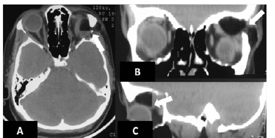
A 33-year-old female patient presented with a swelling and a palpable mass in the supero-temporal region of the left eye. Her past history revealed that her complaints first began at the age of 12 years. Ophthalmological examination revealed a mass on the left upper eyelid. Visual acuity was intact in both eyes, eye movements were free in all directions, and biomicroscopic examination and fundus were seen normal. Laboratory data were within normal limits. An orbital computed tomography (CT) and a magnetic resonance imaging (MRI) were performed with 16-slice multidetector CT and 1.5 Tesla MRI devices, respectively. Orbital CT examination was performed with 0.5 mm sections without contrast material. Orbital MRI examination was performed by obtaining SE T1- weighted(T1W) axial, TSE T2W axial and coronal, and post-contrast T1W axial and coronal images. Intravenous paramagnetic contrast material of 0.1 mmol/kg was administered via antecubital vein for contrast images at the end of the examination. Orbital CT examination revealed a macrolobulated, dumbbell-shaped lesion with a density equal to or less than that of fatty tissue, which had a component that separates zygomatico-frontal suture and extends to subcutaneous fatty tissue at the level of temporal fossa on lateral side. The lesion was mildly compressing the superior rectus muscle and the bulb. The bone window showed separation of orbital lateral zygomatico-frontal suture (Figures 1A-C).
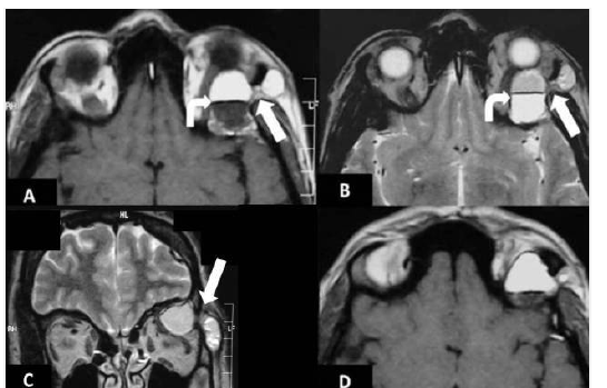
T2W images on MRI examination revealed a hyperintense, dumbbell-shaped, smooth-bordered lesion. T1W axial images showed a hypointense lesion, similar to adjacent tissues. There were two layers in the cyst, with reverse signal between the up and down layers on the T1W and T2W images. This appearance was consistent with a fat-fluid level. Post-contrast T1W axial and coronal images were notable for a mild contrast uptake around the lesion that had a hypointense view (Figures 2 A-D). Based on the CT and MRI findings, the patient was diagnosed with a dermoid cyst. The lesion was excised and the diagnosis of dermoid cyst was confirmed histopathologically.
Discussion
Dermoid cyst is an ectodermal inclusion cyst which may occur anywhere in the body, but its occurrence in orbit is relatively rare. Approximately 50% of tumors that involve the head are found in or adjacent to the orbit (1, 4). The superior temporal quadrant at the zygomatico-frontal suture is the most common location, followed by the nasal aspect of upper orbit at the fronto-ethmoidal suture. They are usually located anteriorly and at paraorbital sites. Dermoid cysts have a slow growth rate. Cysts that are located at upper outer periorbital quadrant may extend deep in the orbita and temporal fossa (dumbbell dermoid), as in our case. Although dumbbell dermoids extend anteriorly as a palpable mass, they may be found in both the orbit and the temporal fossa by destructing lateral orbital wall (5).
Behavioral patterns of dermoid cysts in orbital and periorbital region depend on the location, size, and growth rate of the tumor. These lesions can be examined by dividing them into groups based on deep or superficial location. In these cases, separation of zygomatico-frontal suture may be present and the lesions are usually located on the lateral orbital wall. Zygomatico-frontal suture is located between the zygomatic bone and the zygomatic process of the frontal bone at the level of lateral orbital wall. Normal zygomatico-frontal suture has a zig-zag configuration and contains a thin streak-like fibrous tissue at the center.
Orbital dermoid cysts may cause sequestration on the keratinized surface of the epidermis and extend to infratemporal fossa or infraorbital cavity through the zygomatico-frontal suture. This clinical scenario rarely occurs but it causes zygomatico-frontal suture separation. Lesions with close relationship with deep orbital tissues are also difficult to treat surgically and they may cause complications that lead to injury of orbital structures and optic nerve. Our case also had a dermoid cyst at the lateral orbital wall, which separated zygomatico-frontal suture and compressed deep tissues of the orbital cavity (5–8).
The imaging method to be selected in dermoid cysts depends on the location and the pattern of behavior of the lesion. Orbital CT is superior for demonstration of remodeling or destruction of bones, and presence of a calcific or macroscopic fatty component. Orbital MRI is useful for evaluation of the lesion contents that have a fluid density in CT examination, for demonstration of the relationship with adjacent soft tissues, and intracranial extent of the lesion. Dermoid cysts appear as masses with a well-defined border, a smooth contour, and a low density in orbital CT without contrast. They had a fat density in CT when they contain fatty components. Attenuation level of dermoid cysts ranges between (-60) and (-90) Hounsfield units (HU) in CT. They may contain calcific components. They may show a fat-fluid level. Heterogeneity in the fat tissue consistent with inflammation may be observed adjacent to ruptured dermoid cysts. Rarely, some dermoid cysts may appear uniformly hyperdense in CT examination. Similar to other cysts, dermoid cysts had a low T1W signal and a high T2W signal in orbital MRI if they do not contain any macroscopic fat component. However, depending on the fat component, they show characteristic signal properties of fat tissue when they contain macroscopic fat. These lesions had a signal equal to subcutaneous fatty tissue in all sequences. Postcontrast MRI examination does not show any contrast uptake by the content of the dermoid cysts but cysts may show a thin peripheral contrast uptake. Despite their typical radiological appearances, dermoid cysts may have markedly different sites of location (1, 8–10).
The differential diagnosis of dermoid cysts located in the anterior lateral includes lacrimal gland masses, teratomas and plexiform neuro fibromas. The differential diagnosis of dermoid cysts located in the anterior medial includes both mucoceles and encephaloceles (3, 10, 11). Management of dermoid cysts is surgical with complete excision of the cyst lining, taking care to avoid rupture. All attempts should be made to remove the remnants of the cyst to avoid a potential severe inflammatory reaction (8, 10).
In conclusion, orbital dermoid cysts are common benign lesions. Although their diagnosis and therapy are straightforward, it should be remembered that their clinical signs will be diversified and their surgical treatment complicated due to compressive effects of the deep component. CT and MRI are effective tools for diagnosis of these lesions and determination of their extent.
Declarations
- Author contributions (CRediT)
- All authors contributed equally to all 14 CRediT roles (Conceptualization, Data curation, Formal analysis, Funding acquisition, Investigation, Methodology, Project administration, Resources, Software, Supervision, Validation, Visualization, Writing – original draft, Writing – review & editing).
- Funding
-
Funding information was not reported in the published version of this article.
- Conflicts of interest
- The authors declare that they have no conflict of interest in the current study.
- Ethics approval
- Ethics approval details were not reported in the published version of this article.
- Data availability
- A data availability statement was not reported in the published version of this article.
References (11)
8 of 11 references are linked to their DOI.
- Shawda SJ, Moseley IF. Computed tomography of orbital dermoids: a 20-year review. Clin Radiol. 1999; 54: 821-825 PubMed Google Scholar
- Yeola M, Joharapurkar SR, Bhole AM, Chawla M, Chopra S, Paliwal A. Orbital floor dermoid: an unusual presentation. Indian J Ophthalmol. 2009; 57: 51-52 DOI Crossref PubMed Google Scholar
- Pryor SG, Lewis JE, Weaver AL, Orvidas LJ. Pediatric dermoid cysts of the head and neck. Otolaryngol Head Neck Surg. 2005; 132: 938-942 DOI Crossref PubMed Google Scholar
- Sathananthan N, Moseley IF, Rose GE, Wright JE. The frequency and clinical significance of bone involvement in outer canthus dermoid cysts. Br J Ophthalmol.1993;77:789 -94 DOI Crossref PubMed Google Scholar
- Sowa Y, Nishino K, Numajiri T. Surgical treatment of dumbbell-shaped dermoid cyst extending into the orbit. J Craniofac Surg. 2012; 23: 1198-9 DOI Crossref PubMed Google Scholar
- Samuelson TW, Margo CE, Levy MH, Pusateri TJ. Zygomaticofrontal suture defect associated with orbital dermoid cyst. Surv Ophtalmol. 1988; 33: 127–130 DOI Crossref PubMed Google Scholar
- Emerick GT, Shields CL, Shields JA, Eagle RC Jr, De Potter P, Markowitz GI. Chewing-induced visual impairment from a dumbbell dermoid cyst. Ophthal Plast Reconstr Surg. 1997; 13: 57–61 DOI Crossref PubMed Google Scholar
- Güvenç G, Kızmazoğlu C, Aydın HE, Sayın M, Yüceer N. Orbita İçine Uzanan Dumbbell (Halter) Dermoid Kist ve Cerrahi Tedavisi: Bir Olgu Sunumu. Osmangazi Tıp Dergisi. 2016; 38: 42-7 PubMed Google Scholar
- Rao AA, Naheedy JH, Chen JY, Robbins SL, Ramkumar HL. A clinical update and radiologic review of pediatric orbital and ocular tumors. J Oncol. 2013; 2013: 975908 DOI Crossref PubMed Google Scholar
- Keskinbora K, Hür-Yıldırım H. Orbita ve temporal fossa yerleşimli dermoid kist (Halter kisti). T Klin Oftalmoloji. 2003; 12: 222-6 PubMed Google Scholar
- Chung EM, Murphey MD, Specht CS, Cube R, Smirniotopoulos JG. Pediatric Orbit Tumors and Tumorlike Lesions: Osseous Lesions of the Orbit. Radiographics. 2008; 28: 1193-214 DOI Crossref PubMed Google Scholar
Respond to this article
Comments on a published article are welcome as a Letter to the Editor.
Get an e-mail when new articles in your fields are published
Questions to the authors
Questions are moderated: an editor approves each question, the authors answer, and an editor approves the answer before both appear here.
No answered questions yet.
Ask the authors a question
Discussion
Comments are moderated: they appear after the writer confirms the e-mail address and an editor approves them. Each comment carries a conflict-of-interest statement.
No comments yet.
Write a comment
Read the full text
Open the PDF here
Loading the PDF…
If the PDF does not show here, use the download button above.
How to cite this article
Atalar MH, Ozec AV, Baspinar N. Dumbbell-shaped Dermoid Cyst Extending to Temporal Fossa from Zygomatico-frontal Suture Defect. Ulutas Med J. 2019;5(3):229-232. doi:10.5455/umj.20190706072248
Atalar, M. H., Ozec, A. V., & Baspinar, N. (2019). Dumbbell-shaped Dermoid Cyst Extending to Temporal Fossa from Zygomatico-frontal Suture Defect. The Ulutas Medical Journal, 5(3), 229–232. https://doi.org/10.5455/umj.20190706072248
Atalar, M.H., Ozec, A.V. and Baspinar, N. (2019) 'Dumbbell-shaped Dermoid Cyst Extending to Temporal Fossa from Zygomatico-frontal Suture Defect', The Ulutas Medical Journal, 5(3), pp. 229–232. doi: 10.5455/umj.20190706072248.
@article{umj54732,
  title = {Dumbbell-shaped Dermoid Cyst Extending to Temporal Fossa from Zygomatico-frontal Suture Defect},
  author = {Atalar, Mehmet H and Ozec, Ayse Vural and Baspinar, Nisa},
  journal = {The Ulutas Medical Journal},
  year = {2019},
  volume = {5},
  number = {3},
  pages = {229--232},
  doi = {10.5455/umj.20190706072248},
  url = {https://www.ulutasmedicaljournal.com/?mno=54732},
  issn = {2149-388X}
}