The Ulutas Medical Journal · ISSN 2149-0430 (print), 2149-388X (online) · https://www.ulutasmedicaljournal.com/?mno=21646
Original Article · Open access
Histopathological Examination of Internal Thoracic Artery and Saphenous Vein in Patients with Diabetes Mellitus: Structural Changes in Grafts Used in Bypass Surgery
- Sanko Hast. incilipınar mah. No45 27090 ŞehitkamilGaziantep ROR
Abstract
- Introduction
- Epidemiological studies show that cardiovascular mortality is two to three times higher in male gender and three to five times higher in women with diabetes than those without diabetes. In this study, LIMA, which we frequently use in coronary bypass surgery, and the comparison of structural differences, if any, that may affect the saphenous vein, were analyzed with samples taken from diabetic and non-diabetic patients.
- Materials and Methods
- Twelve diabetic and 12 non-diabetic patients who underwent coronary by-pass between 2003-2004 use of this type were examined. We evaluated the intimal thickness, medial thickness, intima-media ratio, and medial calcification of the biopsies from these grafts used during the operation.
- Results
- LIMA calcification were calculated as 10,84±0,98µ in diabetics and 12±2,15µ in the control (p=0.048). When the calcification in saphenous veins was evaluated as a percentage, it was calculated as 7.19±3,47µ in the diabetic and 7,95±1,16µ in the control (p=0.037). When the LIMA intima thickness was evaluated, it was calculated as 28,75±15,65µ and 26,36±24,05µ in the control (p=0.044). When the LIMA intima-media ratios were examined, it was calculated as 0.128±0.93µ in the diabetic group and 0.117±0.092µ in the control (p=0.041). Measurement of saphenous vein intimal thickness was found as 68,05±47,94µ in the diabetic group and 66,31±48,07µ in the control group (p=0.038). When measured as the medial thickness of the saphenous vein, it was found to be 422,18±131,7µ in the diabetic group and 465,6±146,28µ in the control (p=0.032).
- Conclusion
- We could not find any statistically significant difference between the two groups. Since we could not find a significant difference in terms of grafts between the diabetic and control groups, we can only say that postoperative factors cause premature occlusion of grafts in diabetics.
Keywords
Full text
Introduction
Diabetes is an important cardiovascular risk factor. Epidemiological studies have shown that cardiovascular mortality is two to three times higher in men with diabetes and three to five times higher in women with diabetes (1). Many prospective studies in both type 2 diabetics and non-diabetic individuals have shown that hyperglycemia is an undeniable cardiovascular risk factor. The effect of blood glucose on cardiovascular risk begins well before diabetic values (≥100 mg/dl) and continues to act increasingly with rising glucose values. Findings show that the postprandial blood glucose independently increases the risk of mortality (2). The mechanism by which hyperglycemia leads to macrovascular changes is not fully known. Although the success rate of coronary angioplasty in diabetic patients is similar to those without diabetes, restenosis, and late complication rates were found to be high. Similarly, mortality, early and late mortality, and the need for reoperation after coronary artery bypass (CABG) were found to be higher in diabetic patients compared to those without diabetes (3). In the long-term follow-up and symptoms of patients undergoing CABG, the rate of graft patency used in bypass is important. Besides, the early patency rate depends on the operation technique, the severity of the pathology in the bypassed vessels, and the graft used. While the most ideal graft responds to physiological and anatomical needs, it must provide the necessary conditions for complete revascularization, be long enough to reach the distal coronary vessels, and remain open for life in the patient. Unfortunately, a graft that responds to all these conditions has not been found. The choice of the graft to be used for the operation depends on the maximum advantage that can be tolerated, the patient's age, risk factors, the urgency of the treatment, and finally the surgeon's experience. It became popular when it was reported by Favoloro for the first time that is saphenous vein (GSV, alternately "long saphenous vein) could be used as a graft in occlusive vascular diseases (4). In the early 1970s, when the comparative studies performed with the internal thoracic artery (ITA) showed that saphenous flow was higher, its popularity increased (5). In studies conducted up to our time, 8-12% of saphenous grafts are blocked in the early postoperative period, and at the end of the first year, it has been shown that this rate is 12-20%. After the first year, this occlusion rate drops to 2%. The 5-year occlusion is 22-30% cumulatively (6). After the 5th year, the occlusion rate increases, and 50% of the grafts are blocked in the 10th year (7). In 20 years of follow-up, occlusion has been reported as 81% in the first 5 years, 68% in the 5th-10th year, and 46% in 16-20 years (8). Some of the important factors that determine the patency rate of the saphenous vein are the diameter of the vessel it is made, the amount of injury in the coronary vessel, and the flow rate to the distal. Consecutive by-passes are also found to remain open more than single grafts (9). Endothelial injury leads to the release of the relaxant factor and prostacyclin released from the endothelium, which leads to impairment of endothelial integrity, platelet, and leukocyte adhesion (10). Uniform medial thickening, followed by intimal thickening, may develop in the open graft. Although the pathology has not been clearly explained, it is thought to be due to the migration and proliferation of smooth muscle cells. Intimal thickening may occur with the accumulation of the extracellular matrix or the organization of the mural thrombus (10). However, intimal proliferation is caused by the metamorphosis of endothelial regeneration, the amount of endothelial injury and the effect of platelet-derived growth factor (PDGF) released from the vascular vessel wall (11). Intimal thickening is present in 75% of the grafts at the end of the 1st year and is the most common cause of graft stenosis 6-24 months after the operation (12).
When the saphenous vein is exposed to coronary blood flow during coronary bypass, some hemodynamic changes occur. When the lower extremity veins are exposed to normal arterial pressure, they lose their elasticity and undergo some intrinsic changes to adapt to the arterial pressure. Medial thickening of the vein graft wall and changes in vasomotor functions have been detected in performed angiotensin-induced hypertension experimentally (11).
Increased wall tension is another warning for intimal hyperplasia. A few weeks after implantation, the tension of the artery wall in which it is implanted becomes equal, as a result, smooth muscle cell proliferation slows down (13). After the first year, lipid-loaded atherosclerotic plaque begins to appear (10). Atherosclerosis of the vein graft may develop in the area of intimal hyperplasia or a different region (14). In hyper cholesterol, the pressure applied to the artery during implantation and preparation facilitates lipid collection in the media of the graft (15).
The most commonly used graft in coronary bypass surgery is undoubtedly the Left Internal Thoracic Artery (LIMA). This artery, which is the second branch of the subclavian artery, first attracted attention with publications written in 1972 (16) and became indispensable in the mid- 1980s with the demonstration that the patency rates were over 90% in 12-year follow-up (17). In studies with a large number of patients, it has been found that long-term patency rates are higher than venous grafts, especially in patients with good ventricular function, and higher survival and cardiac events are observed less (18).
It has been reported that smooth muscle cells of venous grafts respond more to growth factors secreted from platelets when compared with the smooth muscle cells of LIMA (19). Although pulsatile mechanical stress is a potent mitogen for the saphenous vein, LIMA is not for smooth muscle cells (20). Prostacyclin, a LIMA vasodilator, and platelet inhibitor secrete higher amounts of venous smooth muscle cells (21). LIMA endothelial cells produce a significantly more potent vasodilator nitric oxide (NO) compared to the saphenous vein (22). For many reasons like this, LIMA has gained the right to be an indispensable graft.
In this study, LIMA, which we frequently use in coronary bypass surgery, and the comparison of structural differences, if any, that may affect the saphenous vein, with samples taken from diabetic and non-diabetic patients. For this purpose, biopsy samples were taken from saphenous vein and LIMA to evaluate intimal thickness, medial thickness, intima-media ratio, medial calcification, which are the markers of atherosclerosis (22). LIMA blood flows were also compared.
Materials and Methods
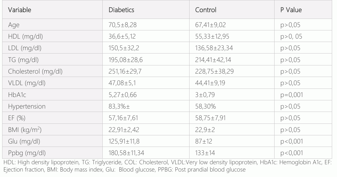
The left internal thoracic artery and saphenous veins of a total of 24 patients who were operated for coronary bypass surgery between 2003-2004 at Gazi University Faculty of Medicine, Department of Cardiovascular Surgery were examined. While 12 of the patients were receiving oral diabetic or insulin therapy for Type-II diabetes, they were operated on by consulting the internal medicine endocrinology clinic and the other 12 patients were admitted as the control (Table-1). Patients whose left ventricular ejection fraction (LVEF) was not lower than 40%, who were diagnosed with coronary disease and decided to coronary bypass by the council jointly made by Cardiology and Cardiovascular surgery clinics, were included in the study. Care was taken to ensure that parameters such as pre-operative information, age, echocardiography findings, coronary angiography findings, drugs used, risk factors, laboratory information were close in the patients.
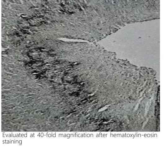
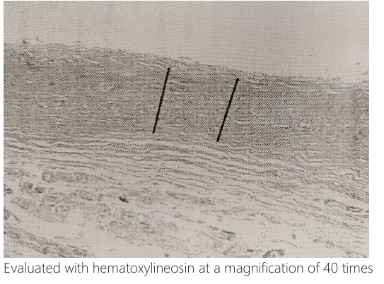
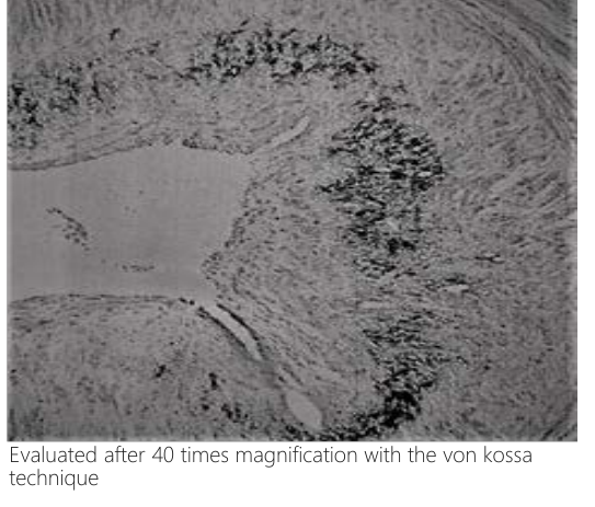
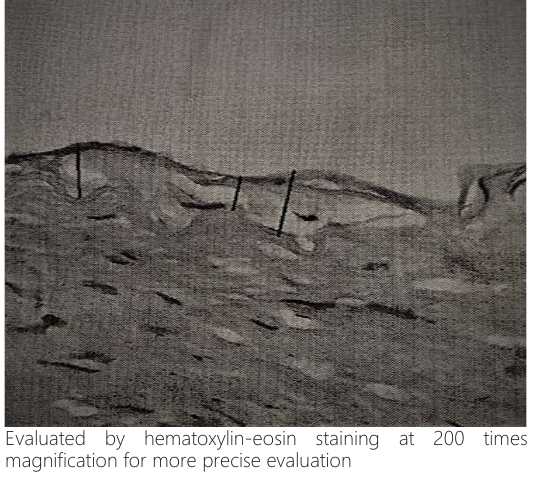
Biopsy materials of 0.5-1 mm from the distal end of LIMA and 1 cm from the proximal ends of the saphenous vein were fixed with formol to be used in the histopathological evaluation of atherosclerosis. It was made by the same team to ensure standardization in LIMA and saphenous vein extraction. Patients' ITA’s were wrapped with papaverine gauze for 2 minutes at 370c, 90-120 mmHg systolic, 60-80mmHg diastolic tension, and then measured by measuring blood flow for 1 minute (ml/min). Biopsy materials were blocked by embedding in paraffin after tissue follow-up. Then, 5 mm sections were prepared. Two sections were taken from each block, one of which was hemotoxylin-eosin, and the other was stained with a technique based on the Von Kossa technique, based on the deposition of silver to phosphate and carbonate compounds in the areas where calcium accumulated and thus showing the calcification. Samba Technologies, Morphometry version 4.32 (2003) France program was used for measurements of intimal thickness, calcification, intima-media ratios, and medial wall thickness. Preparations Images were taken by Olympus BX-51 microscope, Sony 3CCD color video. After measuring intima and media thickness in 5 separate areas from each preparation, the average values were evaluated as a micron (µ).
Statistical Analysis
Statistical analysis was performed using the Statistical Package for the Social Sciences SPSS for Windows 14.0 software. The conformity of the variables to normal distribution was examined using visual (histogram and probability graphs) and analytical methods (Kolmogorov-Smirnov / Shapiro-Wilk tests). In the Kolmogorov-Smirnov test, cases, where the p-value was above 0.05, were accepted as a normal distribution. When a normal distribution was detected, the patient and control group Mann-Whitney U test was used when the student t-test was not performed. A Chi-square test was used to compare the two groups in categorical variables. A significant p-value was accepted as <0.05.
Results
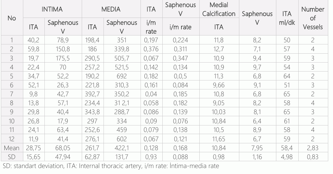
In the evaluations made in the diabetic and control groups, LIMA calcification were calculated as 10,84±0,98 µ percent in diabetics and 12±2,15µ in the control group (p=0.048). When the calcification in saphenous veins was evaluated as a percentage, it was calculated as 7.19±3,47µ in the diabetic group and 7,95±1,16 µ in the control group (p=0.037). When the LIMA intima thickness was evaluated, it was calculated as 28,75±15,65µ and 26,36±24,05µ in the control group (p=0.044). When the LIMA intima-media ratios were examined, it was calculated as 0.128±0.93µ in the diabetic group and 0.117±0.092µ in the control (p=0.041). Measurement of saphenous vein intimal thickness were found as 68,05±47,94µ in the diabetic group and 66,31±48,07µ in the control group (p=0.038).When measured as the medial thickness of the saphenous vein, it was found to be 422,18±131,7µ in the diabetic group and 465,6±146,28µ in the control (p=0.032).
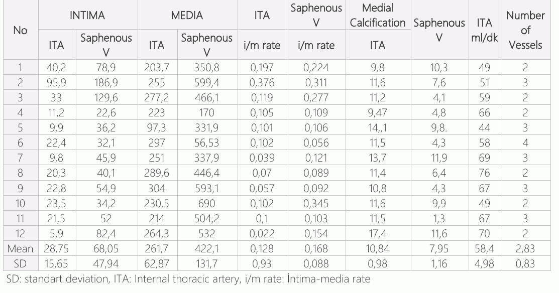
When the saphenous vein intima-media ratio was examined, it was calculated as 0.168±0.088 µ in the diabetic group and 0.165±0.098 µ in the control group (p=0.0385). When the LIMA currents (ml/min) were evaluated, it was 58.41 ±4.98 (ml/min) in the diabetic group versus 60.41±10.23 (ml/min) in the control group (p> 0.039). Comparing the number of bypassed vessels in diabetic group 2,83±0.83 and control 2.58±0,67. Although there were differences between the diabetic and control groups as a result of the examinations, no statistical difference was found.
Discussion
The appropriate graft to be used for coronary bypass in cardiac surgery has led to many studies. While cardiovascular mortality rates tend to decrease in the general population, this rate tends to increase in diabetics (23). Therefore, diabetes has special importance in cardiac surgery. In our study group, 75% of the group was using oral antidiabetic agents (n=9), while 25% were using insulin (n=3). It has been reported in many studies that diabetes is an important risk factor in the development of atherosclerotic lesions and occlusion of bypass grafts, independent of other factors. (24). It has been observed that distal coronary arteries are affected more frequently in diabetics (25). In the BARI study, it was shown that the rate of coronary bypass utilization of diabetic patients depends on the use of LIMA (26). However, bilateral use has been shown to cause an increase in sternal dehiscence and wound infections at a high rate. (27) In addition to studies stating that it is higher in women in terms of mortality (28), there are also studies arguing that there is no difference (29). In terms of age, although there was a difference between the diabetic group (mean 70.5) and the control group (mean 57.31) in our study, no statistically significant difference was observed. In a study with occluded grafts, it is associated with cholesterol (29). The hypercholesterolemia mechanism that initiates, develops, and sustains vein graft atherosclerosis is still not completely explained. Accelerated development of atherosclerosis has been observed due to the increasing pressure in vein grafts. (29). In a study similar to our study, no difference was found in the saphenous vein and LIMA contraction and vasodilatation tests among the grafts used in coronary bypass surgery, but they found that arterial grafts were preserved in histopathological studies (30).
Conclusion
In our literature review, we found that ITA is an indispensable graft in coronary heart surgery, which has been confirmed by many studies and the saphenous vein is used most frequently. In the histopathological examination of our study, we could not find any statistically significant difference between the two groups. At the beginning of this study, we started with the argument that some factors are arising from the grafts themselves in early occlusion. Unfortunately, since we could not find a significant difference in terms of grafts between the diabetic and control groups, we can only say that postoperative factors cause premature occlusion of grafts in diabetics. Prospective studies with larger groups is needed to support this study.
Declarations
- Author contributions (CRediT)
- All authors contributed equally to all 14 CRediT roles (Conceptualization, Data curation, Formal analysis, Funding acquisition, Investigation, Methodology, Project administration, Resources, Software, Supervision, Validation, Visualization, Writing – original draft, Writing – review & editing).
- Funding
-
Funding information was not reported in the published version of this article.
- Conflicts of interest
- The author declared no conflict of interest.
- Ethics approval
- The Gazi University Faculty of Medicine ethics committee approval was obtained for the study.
- Data availability
- A data availability statement was not reported in the published version of this article.
References (30)
22 of 30 references are linked to their DOI.
- Weintraub WS, Wenger KN, Jones LE, Craver MJ, Guyton RA. Changing clinical characteristics of coronary surgery patients; Circulation 1993,88;79-76 Structural Changes in Grafts Used in Bypass Murat Mumtaz Yardımcı PubMed Google Scholar
- Glucose tolerance and mortality: comparison of WHO and American Diabetes Association diagnostic criteria. The DECODE study group. European Diabetes Epidemiology Group. Diabetes Epidemiology: Collaborative analysis Of Diagnostic criteria in Europe. Lancet. 1999:21;354:617-21 DOI Crossref PubMed Google Scholar
- Fontbonne A, Charles MA, Thibult N, Richard JL, Claude JR, et. al. Hyperinsulinaemia as a predictor of coronary heart disease mortality in a healthy population: Paris Prospective Study, 15-year follow-up. Diabetologia 1991;34:356-61 DOI Crossref PubMed Google Scholar
- Detre KM, Lombardero MS, Brooks MM, Hardison RM, Holubkov R, et.al. The effect of previous coronary-artery bypass surgery on the prognosis of patients with diabetes who have acute myocardial infarction. Bypass Angioplasty Revascularization Investigation Investigators, N Engl J Med 2000 6;342:989-97 DOI Crossref PubMed Google Scholar
- Favaloro RG. Saphenous vein autograft replacement of severe segmental coronary artery occlusion: operative technique, Ann Thorac Surg. 1968;5:334-9 DOI Crossref PubMed Google Scholar
- Bartley TD, Bigelow JC, Page US. Aortocoronary bypass grafting with multiple sequential anastomoses to a single vein, Arch Surg. 1972 Dec;105:915-7 DOI Crossref PubMed Google Scholar
- Klima T, Beard EF, Milam JD, MD, Reul GJ, Cooley DA.ş Atherosclerosis of aortacoronarym artery saphenous vein bypass grafts, Cardiovasc Dis. 1979;6:318–23 PubMed Google Scholar
- Fitz Gibbon GM, Leach AJ, Kafka HP. Atherosclerosis of coronary artery bypass grafts and smoking, CMAJ. 1987;136: 45–7 PubMed Google Scholar
- Loop FD, Cosgrove DM, Lytle BW, Thurer RL. Simpfendorfer C, Taylor PC, Proudfit WL. An 11 Year Evolution of Coronary Arterial Surgery (1967-1978), Ann Surg. 1979;190:444–54 DOI Crossref PubMed Google Scholar
- Meeter K,Veldkamp R, Tijssen JGP,Herwerden L,Bos E, Clinical outcome of single versus sequential grafts in coronary bypass operations at ten years’ follow-up, J Thorc. Cardiovasc Surg. 1991;101:1076-81 DOI Crossref PubMed Google Scholar
- Angelini GD, Newby AC. The future of saphenous vein as a coronary artery bypass conduit, Eur Heart J. 1989;10:273-80 DOI Crossref PubMed Google Scholar
- Angelini GD,Bryan AJ,Williams HM,Morgan R,Newby AC, Distention promotes platelet and leukocyte adhesion and reduces short-term patency in pig arteriovenous bypass grafts, J Thorac Cardiovasc Surg. 1990;99:433-9 DOI Crossref PubMed Google Scholar
- Grondin CM, Lespérance J, Bourassa MG, Campeau L. Coronary artery grafting with the saphenous vein or internal mammary artery. Comparison of late results in two consecutive series of patients, Ann Thorac Surg. 1975;20:605- DOI Crossref PubMed Google Scholar
- Ku DD, Caulfield JB, Kirklin JK. Endothelium-dependent responses in long-term human coronary artery bypass grafts, Circulation. 1991;83:402-11 DOI Crossref PubMed Google Scholar
- Quist WC, Haudenschild CC, LoGerfo FW. Qualitative micro scopy of implanted vein grafts. Effects of graft integrity on morphologic fate, J Thorac Cardiovasc Surg. 1992;103:671-7 PubMed Google Scholar
- Markar RS,Kutty R,Edmonds L,Sadat U,Nair S. A meta-analysis of minimally invasive versus traditional open vein harvest technique for coronary artery bypass graft surgery, Interact Cardiovasc Thorac Surg. 2010;10:266-70 DOI Crossref PubMed Google Scholar
- Collaborative overview of randomised trials of antiplatelet therapy: Prevention of death, myocardial infarction, and stroke by prolonged antiplatelet therapy in various categories of patients. Antiplatelet Trialists' Collaboration, BMJ. 1994:8; 308:81–106 DOI Crossref PubMed Google Scholar
- Kirklin JW, Barrat-Boyers BG. Anatomy, dimensions and terminology, Kirklin JW, Barrat-Boyers BG(eds), Cardiac surgery 4 th eddition,p3 Saunders 2012 PubMed Google Scholar
- Okies JE, Bigelow JC, Krause AH, Salomon NW. The left internal mammary artery: The graft choice, Circulation 1984; 213:377-87 PubMed Google Scholar
- Li S, Fan YS, Chow LH, Diepstraten C, Sims SM, et al. Innate diversity of adult human arterial smooth muscle cells, Circulation Research. 2001;89:517–25 DOI Crossref PubMed Google Scholar
- Ko YS, Yeh H, Haw M, Dupont E, Kaba R, et. al. Differential expression of connexin43 and desmin defines two subpopulations of medial smooth muscle cells in the human internal mammary artery, Arteriosclerosis, Thrombosis, and Vascular Biology. 1999;19:1669–80 DOI Crossref PubMed Google Scholar
- Yang Z, Oemar B, Carrel T, Kipfer B, Julmy F, Lüscher T. Different proliferative properties of smooth muscle cells of human arterial and venous bypass vessels, CirculationVolume 97, Issue 2, 20 January 1998, Pages 181-7 DOI Crossref PubMed Google Scholar
- Raggi P, Boulay A, Taber S, Amin N, Dillon M. Cardiac calcification in adult hemodialysis patients, Journal of the American College of Cardiology, 2002;39: 695-01 DOI Crossref PubMed Google Scholar
- Ruengsakulrach P, Sinclair R, Komeda M, Raman J, Gordon I,et. al. Comparative histopathology of radial artery versus ınternal thoracic artery and risk factors for development of intimal hyperplasia and Aatherosclerosis, Circulation volume. 1999;100:139-44 DOI Crossref PubMed Google Scholar
- Chychota N, Gau G, Pluth J, Wallace R, Danielson G. Comparison of operability and surgical results in diabetic and nondiabetic patients, 1973;65:856-62 PubMed Google Scholar
- Calafiore A, Mauro M, Giammarco G, Lorena Iacò A,Canosa C, et. al. Effect of diabetes on early and late survival after isolated first coronary bypass surgery in multivessel disease, J Thorac Cardiovasc Surg. 2003;125:144-54 DOI Crossref PubMed Google Scholar
- Ali WA, Vaidya SR, MD, Ejeh S, Okoroafor K. Meta-analysis study comparing percutaneous coronary intervention/drug eluting stent versus coronary artery bypass surgery of unprotected left main coronary artery disease, Medicine (Baltimore). 2018 DOI Crossref PubMed Google Scholar
- Lee K, Ahn J, Yoon Y, Kang Y, Park S, et.al. Long‐Term (10‐ Year) Outcomes of Stenting or Bypass Surgery for Left Main Coronary Artery Disease in Patients With and Without Diabetes Mellitus, J Am Heart Assoc. 2020;21;9:1-13 Structural Changes in Grafts Used in Bypass Murat Mumtaz Yardımcı DOI Crossref PubMed Google Scholar
- González B, Hernández C, Garza A, Rodríguez V, Hernández C,et. Al. Conduits Used in Coronary Artery Bypass Grafting: A Review of Morphological Studies, Ann Thorac Cardiovasc Surg. 2017;23:55–65 PubMed Google Scholar
- Li F, Sexton K, Hocking K, Osgood M, Eagle S, et. al. Intimal thickness associated with endothelial dysfunction in human vein grafts, J Surg Res. 2013;180:55-62 DOI Crossref PubMed Google Scholar
Read the full text
Open the PDF here
Loading the PDF…
If the PDF does not show here, use the download button above.
How to cite this article
Yardimci MM. Histopathological Examination of Internal Thoracic Artery and Saphenous Vein in Patients with Diabetes Mellitus: Structural Changes in Grafts Used in Bypass Surgery. Ulutas Med J. 2020;6(4):199-207. doi:10.5455/umj.20201030022829
Yardimci, M. M. (2020). Histopathological Examination of Internal Thoracic Artery and Saphenous Vein in Patients with Diabetes Mellitus: Structural Changes in Grafts Used in Bypass Surgery. The Ulutas Medical Journal, 6(4), 199–207. https://doi.org/10.5455/umj.20201030022829
Yardimci, M.M. (2020) 'Histopathological Examination of Internal Thoracic Artery and Saphenous Vein in Patients with Diabetes Mellitus: Structural Changes in Grafts Used in Bypass Surgery', The Ulutas Medical Journal, 6(4), pp. 199–207. doi: 10.5455/umj.20201030022829.